Glycosaminoglycan-based biohybrid hydrogels are highly promising materials for tissue engineering and regenerative medicine due to their ability to provide cell-instructive environments. In this article, Jana Sievers-Liebschner, Ron Dockhorn, Jens Friedrichs, Thomas Kurth, Peter Fratzl, Jens-Uwe Sommer, Carsten Werner, and Uwe Freudenberg investigate the nanoscale molecular network structure of these hydrogels using an integrated analytical approach.
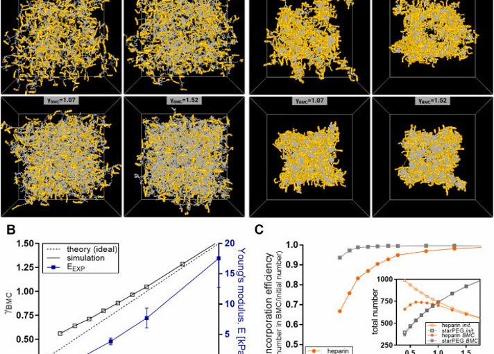
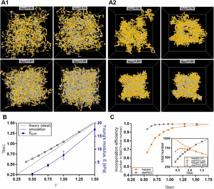
The study combines transmission electron microscopy, X-ray scattering, computer simulations, and AFM-based nanoindentation to quantitatively characterize nanoscale polymer network connectivity and structural inhomogeneities. These parameters are essential for understanding hydrogel mechanics, growth factor delivery, and cell–material interactions relevant to regenerative therapies and organoid culture systems.
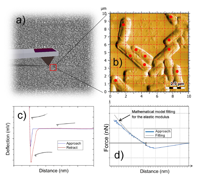
Atomic force microscopy (AFM)-based nanoindentation measurements were performed to determine the mechanical stiffness of the hydrogels in both PBS and ethanol environments. Measurements were conducted using a modified NanoWorld PNP-TR-TL-Au AFM probe equipped with a 10 μm silica bead for colloidal probe nanoindentation.
Nanoindentation experiments were carried out using a set point of 6 nN and an approach/retract velocity of 5 μm/s. At least 70 force–distance curves were recorded for each sample at different positions across the hydrogel surface. Young’s modulus values were extracted using the Hertz model, enabling quantitative evaluation of hydrogel nanomechanical properties.
This work demonstrates how AFM-based nanoindentation with a NanoWorld AFM probe contributes to the detailed characterization of biohybrid hydrogel networks and supports the development of engineered matrices for biomedical applications.
Fig. 5. Computational modelling of starPEG-heparin hydrogel networks. A: Simulation snapshots of hydrated and dehydrated starPEG-heparin hydrogels. For the hydrated network (A1), a good solvent (equivalent to PBS) was assumed. To model the dehydrated state (A2), parameters were adjusted to promote the self-aggregation of starPEG molecules in a poor solvent (e.g., ethanol). Networks with different effective molar ratios after crosslinking (γBMC, where BMC is the Biggest Molecule Cluster) are shown, assuming a 90 % extent of reaction between starPEG (grey) and heparin (yellow). Cube size: L = 150 nm. B: Molar ratio of the BMC γBMC after crosslinking at an extent of reaction p = 0.9, plotted as a function of the initial molar ratio. The dotted line represents the theoretical ideal value, while the blue line shows the experimentally determined Young’s moduli as a function of crosslinking degree. C: Incorporation efficiency of starPEG and heparin within the BMC, calculated as the number of starPEG or heparin molecules in the BMC divided by the number in the reaction mixture (ideal network). Insert: Total number of starPEG or heparin molecules in the reaction mixture (initial) or within the BMC. (For interpretation of the references to colour in this figure legend, the reader is referred to the Web version of this article.)
Full citation:
Sievers-Liebschner, J.; Dockhorn, R.; Friedrichs, J.; Kurth, T.; Fratzl, P.; Sommer, J.-U.; Werner, C.; Freudenberg, U. Unravelling the molecular network structure of biohybrid hydrogels.
Materials Today Bio 2025, 34, 102249. https://doi.org/10.1016/j.mtbio.2025.102249
The surface layer (S-layer) of probiotic bacteria plays an important role in their interaction with the host immune system. In this article, Valentina Taverniti , Paolo D’Incecco , Stefano Farris , Peter Riber Jonsen , Helene Skovsted Eld , Juliane Sørensen, Laura Brunelli, Giacomo Mantegazza, Stefania Arioli and Hanne Frøkiær, investigated how the thickness of the S-layer influences the ability of Lactobacillus helveticus MIMLh5 and Lactobacillus acidophilus NCFM to stimulate Th1-related cytokine production in dendritic cells.
The results revealed an inverse correlation between S-layer thickness and the induction of interleukin-12, indicating that thinner S-layers are associated with a stronger immune-stimulating response. These findings provide new insights into the structure–function relationship of bacterial surface layers and their role in probiotic–host interactions.
Atomic force microscopy (AFM) was used for nanomechanical and morphological characterization of bacterial cells. Measurements were performed using a commercially available AFM instrument operated in contact resonance amplitude imaging (CRAI) mode. An Nanoworld Arrow-FMR force modulation AFM probe was used. This silicon AFM probe features a rectangular beam with a triangular free end and a tetrahedral tip (tip radius ~10 nm, tip height 10–15 μm), a spring constant of 2.8 N/m and a resonance frequency of 75 kHz. Images of 10 × 10 μm² and force–distance curves were recorded at multiple locations on the bacterial surface. Nanomechanical properties, including the elastic (Young’s) modulus, were determined by fitting approach curves to the Hertzian model with an indentation depth set to 2 nm.
Figure S1: Schematic representation of the 4-step procedure for the AFM analysis of the bacteria surface: scanning of the surface in contact resonance amplitude (CRAI) mode (a); creation of the 10-point map of the nanomechanical test (b); generation of the force-distance curves (c); and fitting procedure for the extrapolation of the elastic modulus (d).
Taverniti, V.; D’Incecco, P.; Farris, S.; Jonsen, P. R.; Eld, H. S.; Sørensen, J.; Brunelli, L.; Mantegazza, G.; Arioli, S.; Mora, D.; Guglielmetti, S.; Frøkiær, H. The Capacities of the Probiotic Strains L. helveticus MIMLh5 and L. acidophilus NCFM to Induce Th1-Stimulating Cytokines in Dendritic Cells Are Inversely Correlated with the Thickness of Their S-Layers.
Biomolecules 2025, 15(7), 1012. https://doi.org/10.3390/biom15071012
The article: The Capacities of the Probiotic Strains L. helveticus MIMLh5 and L. acidophilus NCFM to Induce Th1-Stimulating Cytokines in Dendritic Cells Are Inversely Correlated with the Thickness of Their S-Layers, is licensed under a Creative Commons Attribution 4.0 International License, which permits use, sharing, adaptation, distribution and reproduction in any medium or format, as long as you give appropriate credit to the original author(s) and the source, provide a link to the Creative Commons license, and indicate if changes were made. The images or other third party material in this article are included in the article’s Creative Commons license, unless indicated otherwise in a credit line to the material. If material is not included in the article’s Creative Commons license and your intended use is not permitted by statutory regulation or exceeds the permitted use, you will need to obtain permission directly from the copyright holder. To view a copy of this license, visit http://creativecommons.org/licenses/by/4.0/.
The plasticity and growth of plant cell walls (CWs) is still not sufficiently understood on its molecular level. *
Atomic Force Microscopy (AFM) has been shown to be a powerful tool to measure the stiffness of plant tissues. *
In the article “Correlation between plant cell wall stiffening and root extension arrest phenotype in the combined abiotic stress of Fe and Al” Harinderbir Kaur, Jean-Marie Teulon, Christian Godon, Thierry Desnos, Shu-wen W. Chen and Jean-Luc Pellequer describe the use of atomic force microscopy (AFM) to observe elastic responses of the root transition zone of 4-day-old Arabidopsis thaliana wild-type and almt1-mutant seedlings grown under Fe or Al stresses. *
In order to evaluate the relationship between root extension and root cell wall elasticity, the authors used Atomic Force Microscopy to perform vertical indentations on surfaces of living plant roots. *
NanoWorld Pyrex-Nitride silicon-nitride PNP-TR AFM probes with triangular AFM cantilevers were used for the nanoindentation experiments with atomic force microscopy. (PNP-TR AFM cantilever beam 2 (CB2) with a typical force constant of 0.08 N/m and a typical resonant frequency of 17 kHz, typical AFM tip radius 10 nm, macroscopic half cone angles 35°). *
Force-distance (F-D) curves were measured using the Atomic Force Microscope and the PNP-TR AFM tips. *
Because of the heterogeneity of seedling CW surfaces, Harinderbir Kaur et al. used the recently developed trimechanics-3PCS framework for interpreting force-distance curves. The trimechanics-3PCS framework allows the extraction of both stiffness and elasticity along the depth of indentation and permits the investigation of the variation of stiffness with varied depth for biomaterials of heterogeneous elasticity responding to an external force. *
A glass slide with a glued seedling (see Figure 1 cited below) was positioned under the AFM cantilever with the help of an AFM optical camera. Due to the large motorized sample stage of the AFM, the glass slide was adjusted in such a way that the AFM cantilever could be positioned perpendicularly at the longitudinal middle of the glued root. The target working area, the transition zone, was 500 µm away from the root apex, almost twice the length of PNP-TR AFM cantilever. *
As shown in the article the presence of single metal species Fe2+ or Al3+ at 10 μM exerts no noticeable effect on the root growth compared with the control conditions. On the contrary, a mix of both the metal ions produced a strong root-extension arrest concomitant with significant increase of CW stiffness. *
Raising the concentration of either Fe2+or Al3+ to 20 μM, no root-extension arrest was observed; nevertheless, an increase in root stiffness occurred. In the presence of both the metal ions at 10 μM, root-extension arrest was not observed in the almt1 mutant, which substantially abolishes the ability to exude malate. The authors’ results indicate that the combination of Fe2+and Al3+ with exuded malate is crucial for both CW stiffening and root-extension arrest. *
It is shown that the elasticity of plant CW is sensitive and can be used to assess abiotic stresses on plant growth and stiffening. *
However, stiffness increase induced by single Fe2+ or Al3+ is not sufficient for arresting root growth in the described experimental conditions and unexpectedly, the stiffening and the phenotype of seedling roots such as REA are not directly correlated. *
Figure 1 from Harinderbir Kaur et al. 2024 “Correlation between plant cell wall stiffening and root extension arrest phenotype in the combined abiotic stress of Fe and Al”: Principle of nanomechanical measurement of seedling roots with atomic force microscopy. A seedling root (R) is deposited on a microscope slide using silicon glue (N, for Nusil). A fastening band of silicon is seen near the tip of the root (T). The thickness of the fastening band must be thin enough to avoid hindering the AFM support (S), but thick enough to withstand the bending of the root tip. The root is placed under the AFM cantilever (C) as observed by the AFM optical camera. The triangular shaped cantilever (200 µm long) was placed 500 µm away from the root tip in the transition zone where nanoindentation measurements proceeded (as shown). The seedling root and the AFM cantilever are placed within a liquid environment (growth solution, see Supplementary file of the cited article). AFM, atomic force microscopy.
*Harinderbir Kaur, Jean‐Marie Teulon, Christian Godon, Thierry Desnos, Shu‐wen W. Chen and Jean‐Luc Pellequer Correlation between plant cell wall stiffening and root extension arrest phenotype in the combined abiotic stress of Fe and Al
Plant, Cell & Environment 2024; 47:574–584
DOI: https://doi.org/10.1111/pce.14744
The article “Correlation between plant cell wall stiffening and root extension arrest phenotype in the combined abiotic stress of Fe and Al” by Harinderbir Kaur, Jean‐Marie Teulon, Christian Godon, Thierry Desnos, Shu‐wen W. Chen and Jean‐Luc Pellequer is licensed under a Creative Commons Attribution 4.0 International License, which permits use, sharing, adaptation, distribution and reproduction in any medium or format, as long as you give appropriate credit to the original author(s) and the source, provide a link to the Creative Commons license, and indicate if changes were made. The images or other third-party material in this article are included in the article’s Creative Commons license, unless indicated otherwise in a credit line to the material. If material is not included in the article’s Creative Commons license and your intended use is not permitted by statutory regulation or exceeds the permitted use, you will need to obtain permission directly from the copyright holder. To view a copy of this license, visit https://creativecommons.org/licenses/by/4.0/.