
Happy Halloween
In the search for lead-free, Si-microfabrication-compatible piezoelectric materials, thin films of scandium-doped aluminum nitride (Al,Sc)N are of great interest for use in actuators, energy harvesting, and micro-electromechanical-systems (MEMS).*
While the piezoelectric response of AlN increases upon doping with Sc, difficulties are encountered during film preparation because, as bulk solids with completely different structures and large differences in cation radii, ScN (rock salt, cubic) and AlN (wurtzite, hexagonal) are immiscible. *
Consequently, (Al,Sc)N is inherently thermodynamically unstable and prone to phase segregation. Film preparation is further complicated by the technological requirement for polar [001] or [00 1̲] out-of-plane texture, which is achieved using a seeding layer.*
In the article “C-Axis Textured, 2–3 μm Thick Al0.75Sc0.25N Films Grown on Chemically Formed TiN/Ti Seeding Layers for MEMS Applications” Asaf Cohen, Hagai Cohen, Sidney R. Cohen, Sergey Khodorov, Yishay Feldman, Anna Kossoy, Ifat Kaplan-Ashiri, Anatoly Frenkel, Ellen Wachtel, Igor Lubomirsky and David Ehre propose a protocol for successfully depositing [001] textured, 2–3 µm thick films of Al0.75Sc0.25N.*
The procedure relies on the fact that sputtered Ti is [001]-textured α-phase (hcp). Diffusion of nitrogen ions into the α-Ti film during reactive sputtering of Al0.75,Sc0.25N likely forms a [111]-oriented TiN intermediate layer. The lattice mismatch of this very thin film with Al0.75Sc0.25N is ~3.7%, providing excellent conditions for epitaxial growth. In contrast to earlier reports, the Al0.75Sc0.25N films prepared in the current study are Al-terminated. Low growth stress (<100 MPa) allows films up to 3 µm thick to be deposited without loss of orientation or decrease in piezoelectric coefficient. *
An advantage of the proposed technique is that it is compatible with a variety of substrates commonly used for actuators or MEMS, as demonstrated here for both Si wafers and D263 borosilicate glass. Additionally, thicker films can potentially lead to increased piezoelectric stress/strain by supporting application of higher voltage, but without increase in the magnitude of the electric field. *
SEM, AFM, EDS, XRD and XPS techniques were used for the film characterization. For the nanoscale topography maps with atomic force microscopy (AFM) NanoWorld Pyrex-Nitride series PNP-TRS silicon nitride AFM probes were used in peak-force tapping1® mode. *

*Asaf Cohen, Hagai Cohen, Sidney R. Cohen, Sergey Khodorov, Yishay Feldman, Anna Kossoy, Ifat Kaplan-Ashiri, Anatoly Frenkel, Ellen Wachtel, Igor Lubomirsky and David Ehre
C-Axis Textured, 2–3 μm Thick Al0.75Sc0.25N Films Grown on Chemically Formed TiN/Ti Seeding Layers for MEMS Applications
Sensors 2022, 22, 7041
DOI: https://doi.org/10.3390/s22187041
The article “C-Axis Textured, 2–3 μm Thick Al0.75Sc0.25N Films Grown on Chemically Formed TiN/Ti Seeding Layers for MEMS Applications” by Asaf Cohen, Hagai Cohen, Sidney R. Cohen, Sergey Khodorov, Yishay Feldman, Anna Kossoy, Ifat Kaplan-Ashiri, Anatoly Frenkel, Ellen Wachtel, Igor Lubomirsky and David Ehre is licensed under a Creative Commons Attribution 4.0 International License, which permits use, sharing, adaptation, distribution and reproduction in any medium or format, as long as you give appropriate credit to the original author(s) and the source, provide a link to the Creative Commons license, and indicate if changes were made. The images or other third-party material in this article are included in the article’s Creative Commons license, unless indicated otherwise in a credit line to the material. If material is not included in the article’s Creative Commons license and your intended use is not permitted by statutory regulation or exceeds the permitted use, you will need to obtain permission directly from the copyright holder. To view a copy of this license, visit https://creativecommons.org/licenses/by/4.0/.
1Peak Force Tapping® is a registered trademark of Bruker Corporation.
Transient receptor potential (TRP) ion channels are gated by diverse intra- and extracellular stimuli leading to cation inflow (Na+, Ca2+) regulating many cellular processes and initiating organismic somatosensation. *
Structures of most TRP channels have been solved. However, structural and sequence analysis showed that ~30% of the TRP channel sequences, mainly the N- and C-termini, are intrinsically disordered regions (IDRs). Unfortunately, very little is known about IDR ‘structure’, dynamics and function, though it has been shown that they are essential for native channel function. *
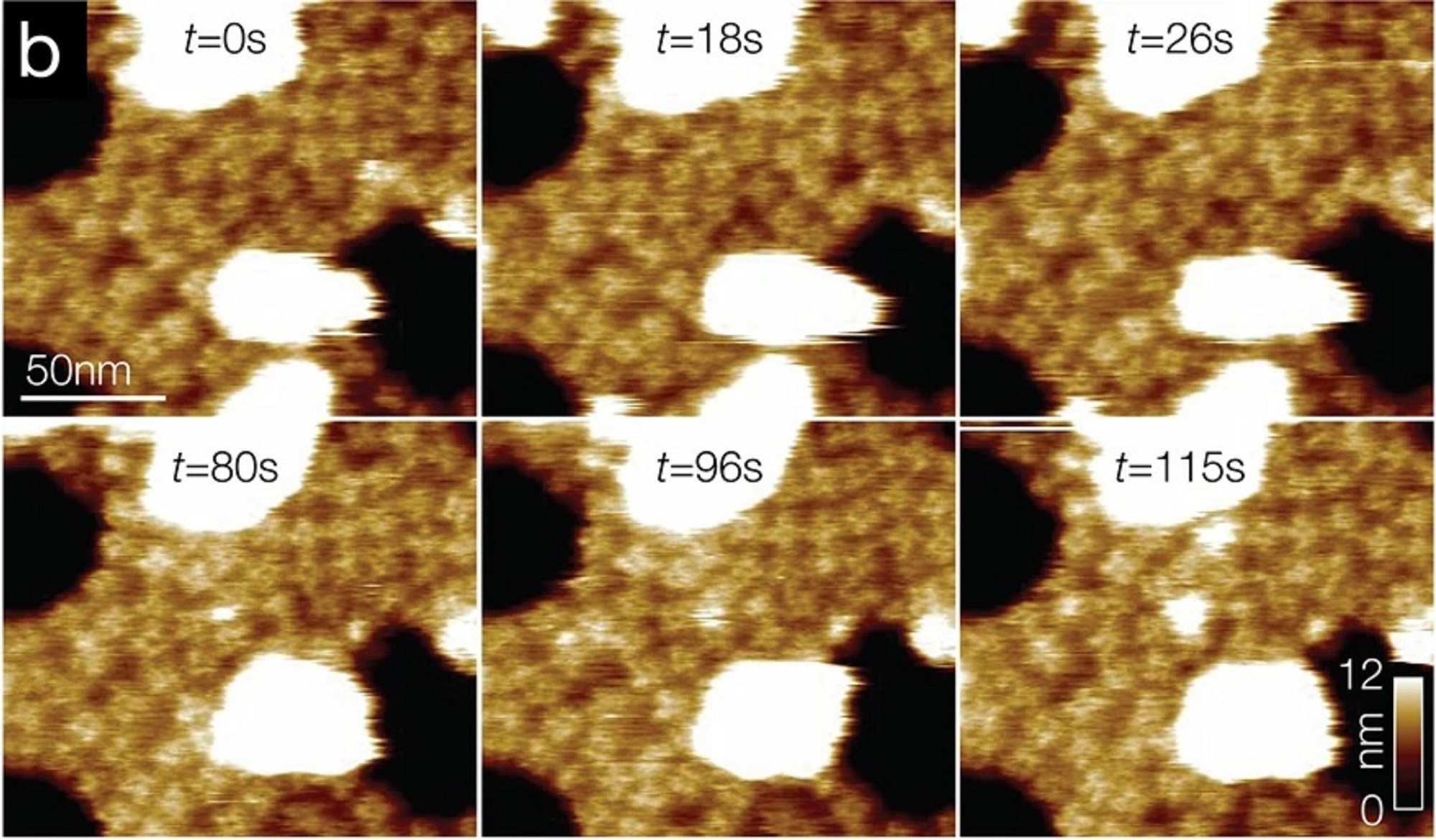
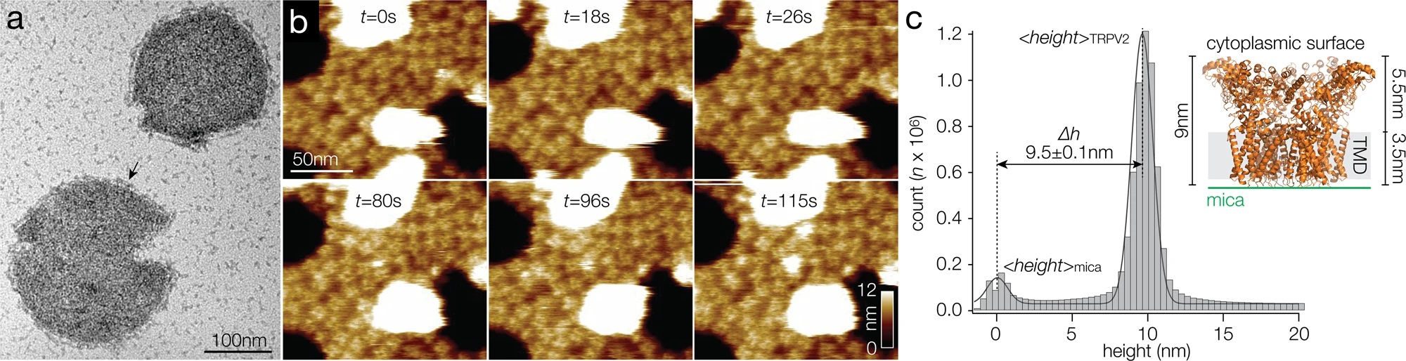
In the article “Intrinsically disordered regions in TRPV2 mediate protein-protein interactions”, Raghavendar R. Sanganna Gari, Grigory Tagiltsev, Ruth A. Pumroy, Yining Jiang, Martin Blackledge, Vera Y. Moiseenkova-Bell and Simon Scheuring imaged TRPV2 channels in membranes using high-speed atomic force microscopy (HS-AFM). *
The dynamic single molecule imaging capability of HS-AFM allowed the authors to visualize IDRs and revealed that N-terminal IDRs were involved in intermolecular interactions. Their work provides evidence about the ‘structure’ of the TRPV2 IDRs, and that the IDRs may mediate protein-protein interactions. *
In total, 1.5 µl of the TRPV2 reconstituted vesicles were deposited on a 1.5-mm2 freshly cleaved mica surface, which was glued with epoxy to the quartz sample stage. After 20–30 min incubation, the sample was gently rinsed with imaging buffer (20 mM Hepes, pH 8.0, 150 mM NaCl) and mounted in the HS-AFM fluid cell. All images in this study were taken using a HS-AFM operated in amplitude modulation mode using optimized scan and feedback parameters and lab-built amplitude detectors and free amplitude stabilizers. *
Short (8 µm) cantilevers (NanoWorld Ultra-Short Cantilevers for High-Speed AFM USC-F1.2-k0.15) with nominal spring constant of 0.15 N/m, resonance frequency of 0.6 MHz, and a quality factor of ∼1.5 in liquid were used. AFM probes were sharpened using oxygen plasma etching to obtain better resolution. *
*Raghavendar R. Sanganna Gari, Grigory Tagiltsev, Ruth A. Pumroy, Yining Jiang, Martin Blackledge, Vera Y. Moiseenkova-Bell and Simon Scheuring
Intrinsically disordered regions in TRPV2 mediate protein-protein interactions
Communications Biology volume 6, Article number: 966 (2023)
DOI: https://doi.org/10.1038/s42003-023-05343-7
Please follow this external link to read the full article: https://rdcu.be/dnNba
The article “Phosphorylation of phase-separated p62 bodies by ULK1 activates a redox-independent stress response” by Raghavendar R. Sanganna Gari, Grigory Tagiltsev, Ruth A. Pumroy, Yining Jiang, Martin Blackledge, Vera Y. Moiseenkova-Bell and Simon Scheuring is licensed under a Creative Commons Attribution 4.0 International License, which permits use, sharing, adaptation, distribution and reproduction in any medium or format, as long as you give appropriate credit to the original author(s) and the source, provide a link to the Creative Commons license, and indicate if changes were made. The images or other third-party material in this article are included in the article’s Creative Commons license, unless indicated otherwise in a credit line to the material. If material is not included in the article’s Creative Commons license and your intended use is not permitted by statutory regulation or exceeds the permitted use, you will need to obtain permission directly from the copyright holder. To view a copy of this license, visit https://creativecommons.org/licenses/by/4.0/.