Layer-stacked gallenene is an emerging two-dimensional material with unique structural and electronic propertiesIn this article, M.Yunusa, A. K.Schulz, T.Parker, et al. investigated the nonlinear optical response of layer-stacked gallenene exhibiting ferroelectric polarization. The material was produced using a liquid metal-based synthesis approach and showed a phase transition associated with its stacked structure.
The authors demonstrated strong second harmonic generation (SHG) signals, revealing the nonlinear optical activity of gallenene and confirming its ferroelectric nature. These findings highlight the potential of gallenene as a novel functional 2D material for advanced optoelectronic and photonic applications.
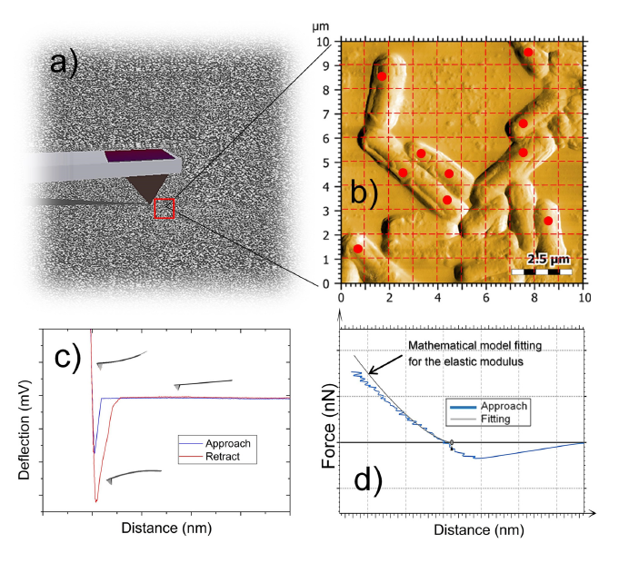
Atomic force microscopy (AFM) was used to characterize transparent lamellar films and helical filaments. Measurements were performed using a commercially available AFM instrument operated in contact mode. A NanoWorld Arrow-CONTR AFM probe with a nominal force constant of 0.2 N/m and a resonance frequency of 14 kHz was used to obtain high-resolution surface morphology data.
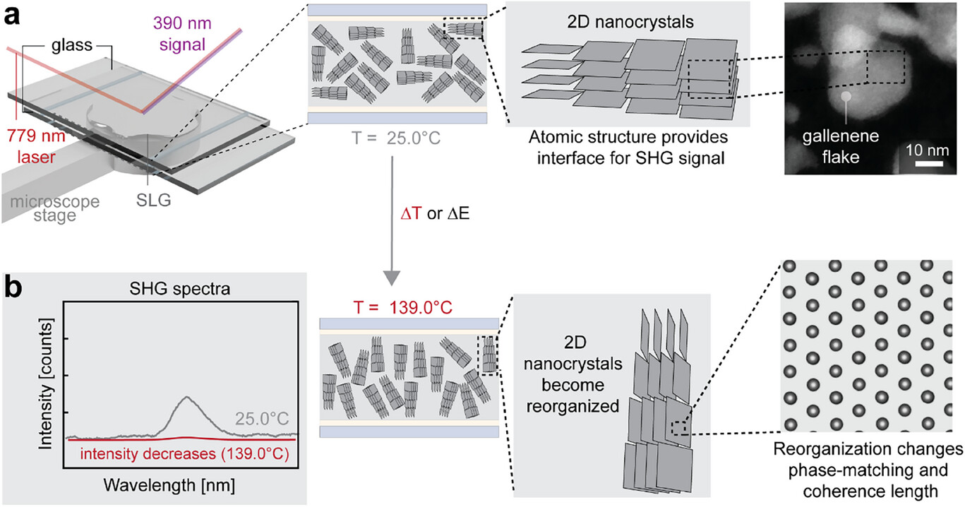
Structure of gallenene and complex anatomy of supercooled liquid gallium. Mechanism for electrical and thermal perturbation. a) Illustration of hypothesized interaction of SHG response with SLG in linearly polarized light showing that thermal perturbations could align the 2D nanocrystals, allowing for an increased SHG medium at either temperature or electrical fields. An example HAADF image of gallenene flake sandwiched between two graphene layers, as depicted in (a) (far right microscope image). b) Structural reorganization of gallenene nanocrystals in the SLG leading to an intensity change in SHG signal as a result of thermal or electrical perturbation.
Full citation:
Yunusa, M.; Schulz, A. K.; Parker, T.; Schneider, F.; Elibol, K.; Predel, M.; Dzíbelová, J.; Rebmann, M.; Gorkan, T.; Ye, J.; Tan, J.-C.; Kang, W.; van Aken, P. A.; Meixner, A. J.; Durgun, E.; Kotakoski, J.; Zhang, D.; Sitti, M. Nonlinear Optical Response in Layer-Stacked Gallenene with Ferroelectric Polarization.
Advanced Materials 2025, 37(44), e01058.
https://doi.org/10.1002/adma.202501058
Attribution 4.0 International By exercising the Licensed Rights (defined below), You accept and agree to be bound by the terms and conditions of this Creative Commons Attribution 4.0 International Public License (“Public License”). To the extent this Public License may be interpreted as a contract, You are granted the Licensed Rights in consideration of Your acceptance of these terms and conditions, and the Licensor grants You such rights in consideration of benefits the Licensor receives from making the Licensed Material available under these terms and conditions. https://creativecommons.org/licenses/by/4.0/