Microfibrillated Cellulose Films from Agri-Food Wastes and Plant Residues for Food Packaging Applications – A Comparative Investigation

Sustainable alternatives to conventional plastic packaging are receiving increasing attention as industries seek circular economy solutions and renewable material sources. In this article, Tommaso Bellesia, Daniele Carullo, Andrea Fachin, Maral Soltanzadeh, Masoud Ghaani, Giorgio Innocenzo Ascrizzi, Laura Piazza, and Stefano Farris investigate the potential of microfibrillated cellulose (MFC) obtained from agri-food waste streams and plant residues as high-performance materials for food packaging applications.
The authors produced MFC dispersions from giant cane, Posidonia oceanica seagrass, and coffee silverskin using high-pressure homogenization and compared the resulting films with a commercially available cellulose-based packaging material. An extensive characterization of the dispersions and films was performed, including rheological, mechanical, optical, barrier, surface, and morphological analyses.
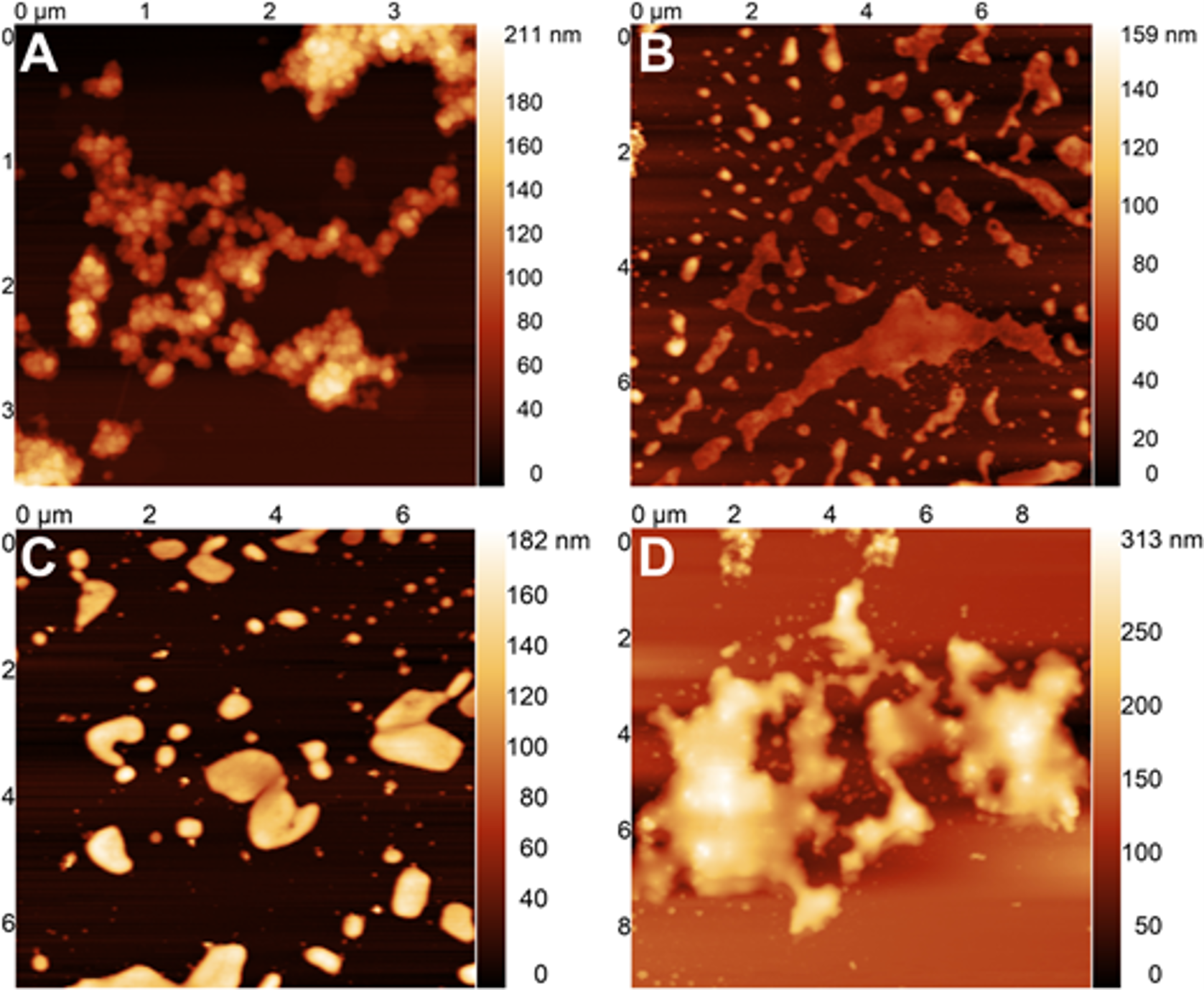
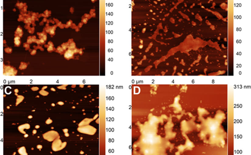
In this article, atomic force microscopy (AFM) confirmed the successful production of microfibrillated cellulose structures with average fibril diameters below 100 nm. The resulting films demonstrated excellent oxygen barrier performance, high stiffness, strong tensile properties, and effective UV-shielding capabilities. Among the investigated materials, films produced from coffee silverskin exhibited particularly promising performance, highlighting the potential of converting agricultural by-products into value-added packaging materials.
To investigate film surface topography, AFM measurements were performed in contact resonance amplitude imaging mode using a NanoWorld Arrow-FMR AFM probe. The AFM probe features a rectangular cantilever with a triangular free end and a tetrahedral tip with a typical radius of curvature of approximately 10 nm. With a spring constant of 2.8 N/m and a resonance frequency of 75 kHz, the Arrow-FMR AFM probe enabled detailed nanoscale characterization of film morphology and surface roughness.
The study demonstrates how AFM analysis using a NanoWorld AFM probe contributes to understanding the relationship between cellulose microstructure and the functional performance of sustainable packaging materials. The results further support the development of renewable, high-performance cellulosic thin films derived from waste biomass sources.


Figure 1Fig. 1. AFM height image of MFC from PO-derived cellulose.

Full citation:
Bellesia, T.; Carullo, D.; Fachin, A.; Soltanzadeh, M.; Ghaani, M.; Ascrizzi, G. I.; Piazza, L.; Farris, S.
Microfibrillated cellulose films from agri-food wastes and plant residues for food packaging applications – A comparative investigation.
Food Packaging and Shelf Life 2026, 54, 101728.
https://doi.org/10.1016/j.fpsl.2026.101728

Attribution 4.0 International
https://creativecommons.org/licenses/by/4.0/

Unravelling the Molecular Network Structure of Biohybrid Hydrogels

Glycosaminoglycan-based biohybrid hydrogels are highly promising materials for tissue engineering and regenerative medicine due to their ability to provide cell-instructive environments. In this article, Jana Sievers-Liebschner, Ron Dockhorn, Jens Friedrichs, Thomas Kurth, Peter Fratzl, Jens-Uwe Sommer, Carsten Werner, and Uwe Freudenberg investigate the nanoscale molecular network structure of these hydrogels using an integrated analytical approach.

The study combines transmission electron microscopy, X-ray scattering, computer simulations, and AFM-based nanoindentation to quantitatively characterize nanoscale polymer network connectivity and structural inhomogeneities. These parameters are essential for understanding hydrogel mechanics, growth factor delivery, and cell–material interactions relevant to regenerative therapies and organoid culture systems.

Atomic force microscopy (AFM)-based nanoindentation measurements were performed to determine the mechanical stiffness of the hydrogels in both PBS and ethanol environments. Measurements were conducted using a modified NanoWorld tipless Pyrex-Nitride  PNP-TR-TL-Au AFM probe equipped with a 10 μm silica bead for colloidal probe nanoindentation.

Nanoindentation experiments were carried out using a set point of 6 nN and an approach/retract velocity of 5 μm/s. At least 70 force–distance curves were recorded for each sample at different positions across the hydrogel surface. Young’s modulus values were extracted using the Hertz model, enabling quantitative evaluation of hydrogel nanomechanical properties.

This work demonstrates how AFM-based nanoindentation with a NanoWorld AFM probe contributes to the detailed characterization of biohybrid hydrogel networks and supports the development of engineered matrices for biomedical applications.


Fig. 5. Computational modelling of starPEG-heparin hydrogel networks
Fig. 5. Computational modelling of starPEG-heparin hydrogel networks. A: Simulation snapshots of hydrated and dehydrated starPEG-heparin hydrogels. For the hydrated network (A1), a good solvent (equivalent to PBS) was assumed. To model the dehydrated state (A2), parameters were adjusted to promote the self-aggregation of starPEG molecules in a poor solvent (e.g., ethanol). Networks with different effective molar ratios after crosslinking (γBMC, where BMC is the Biggest Molecule Cluster) are shown, assuming a 90 % extent of reaction between starPEG (grey) and heparin (yellow). Cube size: L = 150 nm. B: Molar ratio of the BMC γBMC after crosslinking at an extent of reaction p = 0.9, plotted as a function of the initial molar ratio. The dotted line represents the theoretical ideal value, while the blue line shows the experimentally determined Young’s moduli as a function of crosslinking degree. C: Incorporation efficiency of starPEG and heparin within the BMC, calculated as the number of starPEG or heparin molecules in the BMC divided by the number in the reaction mixture (ideal network). Insert: Total number of starPEG or heparin molecules in the reaction mixture (initial) or within the BMC. (For interpretation of the references to colour in this figure legend, the reader is referred to the Web version of this article.)


Full citation:
Sievers-Liebschner, J.; Dockhorn, R.; Friedrichs, J.; Kurth, T.; Fratzl, P.; Sommer, J.-U.; Werner, C.; Freudenberg, U.
Unravelling the molecular network structure of biohybrid hydrogels.
Materials Today Bio 2025, 34, 102249.
https://doi.org/10.1016/j.mtbio.2025.102249

Open Access  The article “ Unravelling the molecular network structure of biohybrid hydrogels”  is licensed under a Creative Commons Attribution 4.0 International License, which permits use, sharing, adaptation, distribution and reproduction in any medium or format, as long as you give appropriate credit to the original author(s) and the source, provide a link to the Creative Commons license, and indicate if changes were made. The images or other third party material in this article are included in the article’s Creative Commons license, unless indicated otherwise in a credit line to the material. If material is not included in the article’s Creative Commons license and your intended use is not permitted by statutory regulation or exceeds the permitted use, you will need to obtain permission directly from the copyright holder. To view a copy of this license, visit http://creativecommons.org/licenses/by/4.0/.

Structural Elucidation of Citric Acid Cross-Linked Pectin and Its Impact on the Properties of Nanocellulose-Reinforced Packaging Films

Citric acid cross-linking is an effective strategy for modifying citrus pectin to enhance its performance in sustainable packaging applications. In this article, Chandra Mohan Chandrasekar, Daniele Carullo, Francesco Saitta, Tommaso Bellesia, Elena Caneva, Chiara Baschieri, Marco Signorelli, Dimitrios Fessas, Stefano Farris and Davide Romano, investigated the structural changes induced by citric acid cross-linking and their influence on the properties of nanocellulose-reinforced packaging films..

The authors demonstrated that cross-linking significantly alters the structure–property relationship of the biopolymer matrix, leading to improved film integrity and modified surface morphology. These results provide valuable insight into biopolymer modification strategies for the development of environmentally friendly packaging materials.

Atomic force microscopy (AFM) was employed to characterize the surface morphology of the films. Measurements were performed using a commercially available AFM instrument operated in contact resonance amplitude imaging (CRAI) mode. A NanoWorld Arrow-FMR AFM probe was used.

This AFM probe features a rectangular beam with a triangular free end and a tetrahedral tip (tip radius ~10 nm, tip height 10–15 μm), with a spring constant of 2.8 N/m and a resonance frequency of 75 kHz. Root mean square surface roughness values were calculated from at least five height-mode images.

Fig. 3. 2D synchronized correlation analysis of FTIR spectra for CLCP packaging film trials.
Fig. 3. 2D synchronized correlation analysis of FTIR spectra for CLCP packaging film trials. [The intensity of the auto-peaks on the diagonal line represents the overall change in spectral intensity. The key region of interest is the strong auto-peak around 1700 cm−1, highlighted by

Full citation: Chandrasekar, C. M.; Carullo, D.; Saitta, F.; Bellesia, T.; Caneva, E.; Baschieri, C.; Signorelli, M.; Fessas, D.; Farris, S.; Romano, D.
Structural elucidation of citric acid cross-linked pectin and its impact on the properties of nanocellulose-reinforced packaging films.
International Journal of Biological Macromolecules 2025, 333(2), 148869. https://doi.org/10.1016/j.ijbiomac.2025.148869

Attribution 4.0 International By exercising the Licensed Rights (defined below), You accept and agree to be bound by the terms and conditions of this Creative Commons Attribution 4.0 International Public License (“Public License”). To the extent this Public License may be interpreted as a contract, You are granted the Licensed Rights in consideration of Your acceptance of these terms and conditions, and the Licensor grants You such rights in consideration of benefits the Licensor receives from making the Licensed Material available under these terms and conditions. https://creativecommons.org/licenses/by/4.0/legalcode.en