Rapid changes in tissue mechanics regulate cell behaviour in the developing embryonic brain

In their short report “Rapid changes in tissue mechanics regulate cell behaviour in the developing embryonic brain” published in January 2019, Amelia J Thompson, Eva K Pillai, Ivan B Dimov, Sarah K Foster, Christine E Holt, and Kristian Franze describe how they used time-lapse in vivo atomic force microscopy (tiv-AFM), a method that combines sensitive upright epi-fluorescence imaging of opaque samples, with iterated AFM indentation measurements of in vivo tissue at cellular resolution and at a time scale of tens of minutes, in order to enable time-resolved measurements of developmental tissue mechanics.*

The technique developed by Thompson, Pillai et al. is a useful tool that can help elucidate how variations in stiffness control the brain wiring process. It could also be used to look into how other developmental or regenerative processes, such as the way neurons reconnect after injuries to thebrain or spinal cord, may be regulated by mechanical tissue properties.*

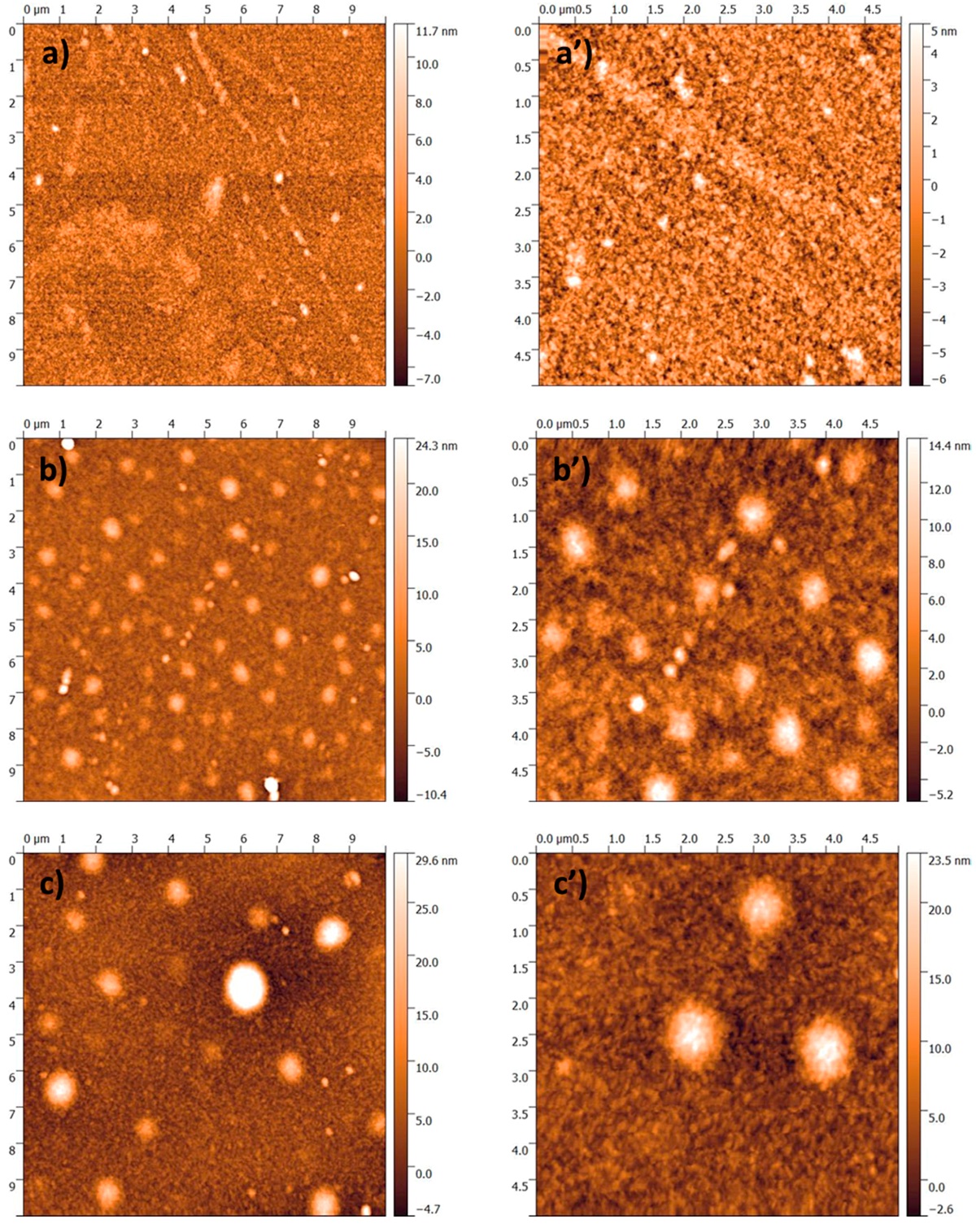
NanoWorld Arrow-TL1 tipless cantilevers were used for the AFM-based stiffness measurements. (Monodisperse spherical polystyrene beads were glued to the cantilever ends as probes.)

NanoWorld Arrow-TL1 tipless cantilever for atomic force microscopy
NanoWorld Arrow-TL1 tipless AFM cantilever

*Amelia J Thompson, Eva K Pillai, Ivan B Dimov, Sarah K Foster, Christine E Holt, Kristian Franze
Rapid changes in tissue mechanics regulate cell behaviour in the developing embryonic brain
eLife 2019; 8:e39356
DOI: https://doi.org/10.7554/eLife.39356

Please follow this external link to the full article: https://cdn.elifesciences.org/articles/39356/elife-39356-v1.pdf

Open Access: The article « Rapid changes in tissue mechanics regulate cell behaviour in the developing embryonic brain » by Amelia J Thompson et al. is licensed under a Creative Commons Attribution 4.0 International License, which permits use, sharing, adaptation, distribution and reproduction in any medium or format, as long as you give appropriate credit to the original author(s) and the source, provide a link to the Creative Commons license, and indicate if changes were made. The images or other third party material in this article are included in the article’s Creative Commons license, unless indicated otherwise in a credit line to the material. If material is not included in the article’s Creative Commons license and your intended use is not permitted by statutory regulation or exceeds the permitted use, you will need to obtain permission directly from the copyright holder. To view a copy of this license, visit http://creativecommons.org/licenses/by/4.0/.


New Insights in the Ion Beam Sputtering Deposition of ZnO-Fluoropolymer Nanocomposites

Surface modification treatments able to confer antistain/antibacterial properties to natural or synthetic materials are receiving increasing attention among scientists. Ion beam co-sputtering (IBS) of zinc oxide (ZnO) and poly-tetrafluoroethylene (PTFE) targets allows for the preparation of novel multifunctional coatings composed of antimicrobial ZnO nanoparticles (NPs) finely dispersed in an antistain PTFE polymeric matrix.*

In the article “New Insights in the Ion Beam Sputtering Deposition of ZnO-Fluoropolymer Nanocomposites” Maria Chiara Sportelli, Marco Valentini, Rosaria Anna Picca, Antonella Milella, Angelo Nacci, Antonio Valentini and Nicola Cioffi describe the use of X-ray photoelectron spectroscopy (XPS), atomic force microscopy (AFM), and transmission electron microscopy (TEM) for the characterization of the IBS deposited coatings in order to obtain information on the materials’ surface composition, with deep insight into the nanocoatings’ morphology as a function of the ZnONP loadings.*

The AFM micrographs shown in this article were acquired on 150-nm-thick films in dynamic (“tapping”) mode, in air, using NanoWorld Pointprobe® NCL AFM probes.

Figure 2 from “New Insights in the Ion Beam Sputtering Deposition of ZnO-Fluoropolymer Nanocomposites” by Maria Chiara Sportelli et al.: Atomic force microscopy (AFM) micrographs of ZnO-CFx nanocomposites having an inorganic phase volume fraction of φ = 0.05 (a–a’), φ = 0.10 (b–b’), and φ = 0.15 (c–c’). NanoWorld Pointprobe® NCL AFM probes were used.
Figure 2 from “New Insights in the Ion Beam Sputtering Deposition of ZnO-Fluoropolymer Nanocomposites” by Maria Chiara Sportelli et al.: Atomic force microscopy (AFM) micrographs of ZnO-CFx nanocomposites having an inorganic phase volume fraction of φ = 0.05 (a–a’), φ = 0.10 (b–b’), and φ = 0.15 (c–c’).

*Maria Chiara Sportelli, Marco Valentini, Rosaria Anna Picca, Antonella Milella, Angelo Nacci, Antonio Valentini and Nicola Cioffi
New Insights in the Ion Beam Sputtering Deposition of ZnO-Fluoropolymer Nanocomposites
Applied Sciences 2018, 8(1), 77
DOI: 10.3390/app8010077

Please follow this external link for the full article: https://www.mdpi.com/2076-3417/8/1/77/htm

Open Access: The article « New Insights in the Ion Beam Sputtering Deposition of ZnO-Fluoropolymer Nanocomposites » by Maria Chiara Sportelli et al. is licensed under a Creative Commons Attribution 4.0 International License, which permits use, sharing, adaptation, distribution and reproduction in any medium or format, as long as you give appropriate credit to the original author(s) and the source, provide a link to the Creative Commons license, and indicate if changes were made. The images or other third party material in this article are included in the article’s Creative Commons license, unless indicated otherwise in a credit line to the material. If material is not included in the article’s Creative Commons license and your intended use is not permitted by statutory regulation or exceeds the permitted use, you will need to obtain permission directly from the copyright holder. To view a copy of this license, visit http://creativecommons.org/licenses/by/4.0/.

Magnetization-polarization cross-control near room temperature in hexaferrite single crystals

In their publication “Magnetization-polarization cross-control near room temperature in hexaferrite single crystals” V. Kocsis, T. Nakajima, M. Matsuda, A. Kikkawa, Y. Kaneko, J. Takashima, K. Kakurai, T. Arima, F. Kagawa, Y. Tokunaga, Y. Tokura and Y. Taguchi report that they have successfully stabilized a simultaneously ferrimagnetic and ferroelectric phase in a Y-type hexaferrite single crystal up to 450 K, and demonstrated the reversal of large non-volatile M by E field close to room temperature. Manipulation of the magnetic domains by E field was directly visualized at room temperature by using magnetic force microscopy.*

NanoWorld MFMR AFM probes with a hard magnetic coating were used for the magnetic force microscopy measurements described in this article.

Figure 5 from “Magnetization-polarization cross-control near room temperature in hexaferrite single crystals” by V. Kocsis et al.: Real-space magnetic force microscopy (MFM) images. The MFM images were taken on the same 10 × 10 μm2 region of a BSCFAO crystal with an ac face (see Supplementary Figs. 3, 9 and 10) at room temperature. Prior to the MFM measurements, the sample was poled to a single-domain ME state using (+E0, +H0) poling fields in a E ⊥ H; E, H ⊥ c configuration. Panel a shows the changes in the magnetic domain pattern caused by two successive applications of the E field with different signs (the initial state is labeled as the 0th). The images include small regions, R1 and R2, where two representative cases of DW motion are observed. Around R1, the negatively magnetized domain (denoted with blue color, MFM phase shift Δφ < 0) expands and shrinks along the c-axis upon the first and second applications of E-field, respectively. On the other hand, around R2, a positively magnetized domain (denoted with red color, Δφ > 0) is pushed into the view area from the upper side along the ab plane. These two cases are further displayed as line profiles of the MFM phase shift (Δφ) data along the b A−A′ and c B−B′ lines. Panels d, e show the schematic illustration of these two cases of domain wall motions for the second E-field switch, respectively. NanoWorld MFMR AFM probes were used for the magnetic force microscopy.
Figure 5 from “Magnetization-polarization cross-control near room temperature in hexaferrite single crystals” by V. Kocsis et al.: Real-space magnetic force microscopy (MFM) images. The MFM images were taken on the same 10 × 10 μm2 region of a BSCFAO crystal with an ac face (see Supplementary Figs. 3, 9 and 10) at room temperature. Prior to the MFM measurements, the sample was poled to a single-domain ME state using (+E0, +H0) poling fields in a E ⊥ H; E, H ⊥ c configuration. Panel a shows the changes in the magnetic domain pattern caused by two successive applications of the E field with different signs (the initial state is labeled as the 0th). The images include small regions, R1 and R2, where two representative cases of DW motion are observed. Around R1, the negatively magnetized domain (denoted with blue color, MFM phase shift Δφ < 0) expands and shrinks along the c-axis upon the first and second applications of E-field, respectively. On the other hand, around R2, a positively magnetized domain (denoted with red color, Δφ > 0) is pushed into the view area from the upper side along the ab plane. These two cases are further displayed as line profiles of the MFM phase shift (Δφ) data along the b A−A′ and c B−B′ lines. Panels d, e show the schematic illustration of these two cases of domain wall motions for the second E-field switch, respectively.

*V. Kocsis, T. Nakajima, M. Matsuda, A. Kikkawa, Y. Kaneko, J. Takashima, K. Kakurai, T. Arima, F. Kagawa, Y. Tokunaga, Y. Tokura, Y. Taguchi
Magnetization-polarization cross-control near room temperature in hexaferrite single crystals
Nature Communications, volume 10, Article number: 1247 (2019)
DOI: https://doi.org/10.1038/s41467-019-09205-x

Please follow this external link to the full article: https://www.nature.com/articles/s41467-019-09205-x

Open Access The article ” Magnetization-polarization cross-control near room temperature in hexaferrite single crystals” by V. Kocsis et al. is licensed under a Creative Commons Attribution 4.0 International License, which permits use, sharing, adaptation, distribution and reproduction in any medium or format, as long as you give appropriate credit to the original author(s) and the source, provide a link to the Creative Commons license, and indicate if changes were made. The images or other third party material in this article are included in the article’s Creative Commons license, unless indicated otherwise in a credit line to the material. If material is not included in the article’s Creative Commons license and your intended use is not permitted by statutory regulation or exceeds the permitted use, you will need to obtain permission directly from the copyright holder. To view a copy of this license, visit http://creativecommons.org/licenses/by/4.0/.