Composite
carbon nanofibres (CNFs) are highly interesting materials which are usable in a
wide array of applications e.g. electrode materials for biosensors, lithium ion
batteries, fuel cells and supercapacitors.*
In their paper
“Electrical conductivity of silver nanoparticle doped carbon nanofibres
measured by CS-AFM” Wael Ali, Valbone Shabani, Matthias Linke,
Sezin Sayin, Beate Gebert, Sedakat Altinpinar, Marcus Hildebrandt, Jochen S.
Gutmann and Thomas Mayer-Gall present a study on the electrical properties of
composite carbon nanofibres (CNFs) using current-sensitive atomic force
microscopy (CS-AFM).*
This technique makes it possible to explore the electrical properties of single fibers and hence derive relationships between the structural features and the electrical properties. NanoWorld AFM probes with conductive PtIr5 coated silicon tips (force constant 2.8 N m−1, length 240 μm, mean width 35 μm and a thickness of 3 μm, and tip height 10–15 μm) Arrow-EFM were used.*
The results presented in the paper show that the composite CNFs have a higher electrical conductivity than the neat CNFs and both the average diameter of the fibers and the electrical conductivity increase with an increasing AgNP content.*
Fig. 8 from “Electrical conductivity of silver nanoparticle doped carbon nanofibres measured by CS-AFM “ by Wael Ali et al.: CS-AFM analysis of CNFs processed from PAN nanofibres electrospun with different concentrations. Images show the friction and current after both stabilisation (a) and carbonisation (b) processes. The applied bias voltage was +0.15 V. The scan area was 5 × 5 μm2 with a scale bar of 1 μm.
*Wael Ali,
Valbone Shabani, Matthias Linke, Sezin Sayin, Beate Gebert, Sedakat Altinpinar,
Marcus Hildebrandt, Jochen S. Gutmann, Thomas Mayer-Gall Electrical conductivity of silver
nanoparticle doped carbon nanofibres measured by CS-AFM
RSC Adv., 2019, 9, 4553-4562
DOI: 10.1039/C8RA04594A
Open Access: The article “Electrical conductivity of silver nanoparticle doped carbon nanofibres measured by CS-AFM” by Wael Ali, Valbone Shabani, Matthias Linke, Sezin Sayin, Beate Gebert, Sedakat Altinpinar, Marcus Hildebrandt, Jochen S. Gutmann and Thomas Mayer-Gall is licensed under a Creative Commons Attribution 3.0 International License, which permits use, sharing, adaptation, distribution and reproduction in any medium or format, as long as you give appropriate credit to the original author(s) and the source, provide a link to the Creative Commons license, and indicate if changes were made. To view a copy of this license, visit https://creativecommons.org/licenses/by/3.0/.
Mucosal immunoglobulins comprise mainly secretory IgA antibodies (SIgAs), which are the major contributor to pathogen-specific immune responses in mucosal tissues. SIgAs exist as mainly dimers and tetramers and play critical roles in mucosal immune responses against influenza.*
Detailed characterization of these anti-viral SIgA is important for better understanding of the mechanisms underlying anti-viral immunity.* In their article “IgA tetramerization improves target breadth but not peak potency of functionality of anti-influenza virus broadly neutralizing antibody” Saito S, Sano K, Suzuki T, Ainai A, Taga Y, Ueno T, et al. (2019) describe a means of generating a recombinant tetrameric monoclonal SIgA to enable exhaustive characterization of tetrameric SIgAs. The tetrameric monoclonal SIgA possessing variable regions of anti-influenza viruses broadly neutralizing antibody show that tetramerization of SIgA improves target breadth, but not the peak potency, of their anti-viral functions.* These results broaden the knowledge about the fundamental role of SIgA tetramerization in anti-viral humoral response at the human respiratory mucosa.*
The high speed atomic force microscopy ( HS-AFM ) experiments mentioned in the article were performed using a NanoWorld Ultra-Short Cantilever USC-F1.2-k0.15.
Fig 1. Production of recombinant tetrameric monoclonal SIgAs from ” IgA tetramerization improves target breadth but not peak potency of functionality of anti-influenza virus broadly neutralizing antibody ” by Saito S, Sano K, Suzuki T, Ainai A, Taga Y, Ueno T, et al. (2019) :
(A) Recombinant monoclonal IgA antibodies purified from the culture supernatant of cells co-transfected with A1+L (left upper), A1+L+J (left lower), A1+L+J+SC (right upper), or A2m2+L+J+SC (right lower), were subjected to size exclusion chromatography (SEC) analysis. A chromatogram showing absorbance at 280 nm revealed three major peaks: peak A (retention volume around 10.4 ml), peak B (retention volume around 9.3 ml), and peak C (retention volume around 8.4 ml). Data are representative of three independent experiments. (B) SDS-PAGE and BN-PAGE analysis of IgG and IgA1/IgA2m2 in each peak fraction (peak A, B, and C) purified from cells co-expressing SC (A1+L+J+SC or A2m2+L+J+SC). (C, D, E) High-mass MALDI-TOF MS analysis of the each peak fraction containing recombinant IgA1 purified from the culture supernatant of cells transfected with A1, L, J, and SC. (C) One main peak (arrow) corresponding to monomer (Mo) was detected in the peak A fraction. (D) Two main peaks (arrows) corresponding to a dimer (Di) and a di-cation dimer (Di2+) were detected in the peak B fraction. (E) Three main peaks (arrows) corresponding to a tetramer (Te), trimer (Tr), and di-cation tetramer (Te2+) were detected in the peak C fraction. (F, G) High-mass MALDI-TOF MS analysis of the each peak fraction of recombinant IgA2m2 purified from the culture supernatant from cells transfected with A2m2, L, J, and SC. (F) One main peak (arrow) corresponding to a monomer (Mo) was detected in the peak A fraction. (G) Three main peaks (arrows) corresponding to a tetramer (Te), a trimer (Tr), and a di-cation tetramer (Te2+) were detected in the peak C fraction. (H) Quantification of the amount of each subunit within the peak B or C fraction of recombinant SIgA1 or SIgA2m2 antibodies purified from the culture supernatant of cells transfected with A1/L/J,/SC or A2m2/L/J/SC using LC-MS with stable isotope-labeled standard peptides. The abundance of each subunit to that of J chain is expressed as a ratio. Data are expressed as box-and-whisker plot with minimum, maximum, median, upper and lower quartiles (n = 6–7). (I) HS-AFM image of peak C derived from a recombinant SIgA1 (A1Te) or SIgA2m2 (A2m2Te) antibody purified from the culture supernatant of cells transfected with A1/L/J/SC or A2m2/L/J/SC. Scale bar, 20 nm.
Open Access: The article « IgA tetramerization improves target breadth but not peak potency of functionality of anti-influenza virus broadly neutralizing antibody » by Saito S, Sano K, Suzuki T, Ainai A, Taga Y, Ueno T, et al. (2019) is licensed under a Creative Commons Attribution 4.0 International License, which permits use, sharing, adaptation, distribution and reproduction in any medium or format, as long as you give appropriate credit to the original author(s) and the source, provide a link to the Creative Commons license, and indicate if changes were made. The images or other third party material in this article are included in the article’s Creative Commons license, unless indicated otherwise in a credit line to the material. If material is not included in the article’s Creative Commons license and your intended use is not permitted by statutory regulation or exceeds the permitted use, you will need to obtain permission directly from the copyright holder. To view a copy of this license, visit http://creativecommons.org/licenses/by/4.0/.
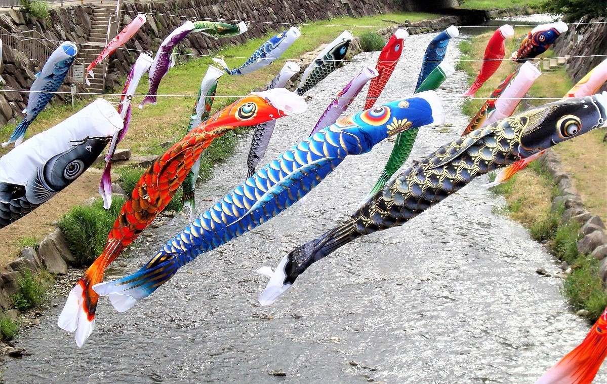
Today is Children’s Day in Japan and many mulit-colored carp-shaped koinobori streamers will flutter in the wind.
So it is the perfect day to share the publication “Piezoelectricity of green carp scales” by Y. Jiang et al. with you.
Piezoelectricity takes part in multiple important functions and processes in biomaterials often vital to the survival of organisms. In their publication , “Piezoelectricity of green carp scales” Y. Jiang et al. investigate the piezoelectric properties of fish scales of green carp by directly examining their morphology at nanometer levels. From the clear distinctions between the composition of the inner and outer surfaces of the scales that could be found, the authors identified the piezoelectricity to originate from the presence of hydroxyapatite which only exists on the surface of the fish scales.*
koinobori – carp streamers in Matsumoto Japan
These findings reveal a different mechanism of how green carp are sensitive to their surroundings and should be helpful to studies related to the electromechanical properties of marine life and the development of bio-inspired materials. As easily accessible natural polymers, fish scales can be employed as highly sensitive piezoelectric materials in high sensitive and high speed devices as well as be exploited for invasive diagnostics and other biomedical implications.*
For the harmonic responses of both 1st order and 2nd order described in this publication, NanoWorld Arrow-CONTPt AFM probes were used.
FIG. 6 from “Piezoelectricity of green carp scales “ by H. Y. Jiang et al.: First and second harmonic responses of (a) domain I and (b) domain IV. The straight line fitting for the amplitude of first harmonic response of (c) domain I and (d) domain IV by applying a series of bias.
*Y. Jiang, F. Yen, C. W. Huang, R. B. Mei, and L. Chen Piezoelectricity of green carp scales
AIP Advances 7, 045215 (2017)
DOI: https://doi.org/10.1063/1.4979503
Open Access The article “Piezoelectricity of green carp scales” by Y. Jiang, F. Yen, C. W. Huang, R. B. Mei and L. Chen is licensed under a Creative Commons Attribution 4.0 International License, which permits use, sharing, adaptation, distribution and reproduction in any medium or format, as long as you give appropriate credit to the original author(s) and the source, provide a link to the Creative Commons license, and indicate if changes were made. The images or other third party material in this article are included in the article’s Creative Commons license, unless indicated otherwise in a credit line to the material. If material is not included in the article’s Creative Commons license and your intended use is not permitted by statutory regulation or exceeds the permitted use, you will need to obtain permission directly from the copyright holder. To view a copy of this license, visit http://creativecommons.org/licenses/by/4.0/.