Sustainable alternatives to conventional plastic packaging are receiving increasing attention as industries seek circular economy solutions and renewable material sources. In this article, Tommaso Bellesia, Daniele Carullo, Andrea Fachin, Maral Soltanzadeh, Masoud Ghaani, Giorgio Innocenzo Ascrizzi, Laura Piazza, and Stefano Farris investigate the potential of microfibrillated cellulose (MFC) obtained from agri-food waste streams and plant residues as high-performance materials for food packaging applications.
The authors produced MFC dispersions from giant cane, Posidonia oceanica seagrass, and coffee silverskin using high-pressure homogenization and compared the resulting films with a commercially available cellulose-based packaging material. An extensive characterization of the dispersions and films was performed, including rheological, mechanical, optical, barrier, surface, and morphological analyses.
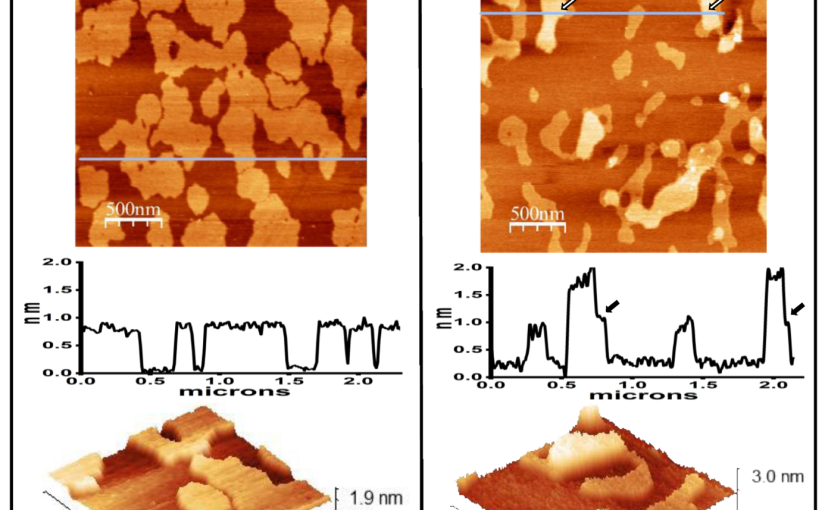
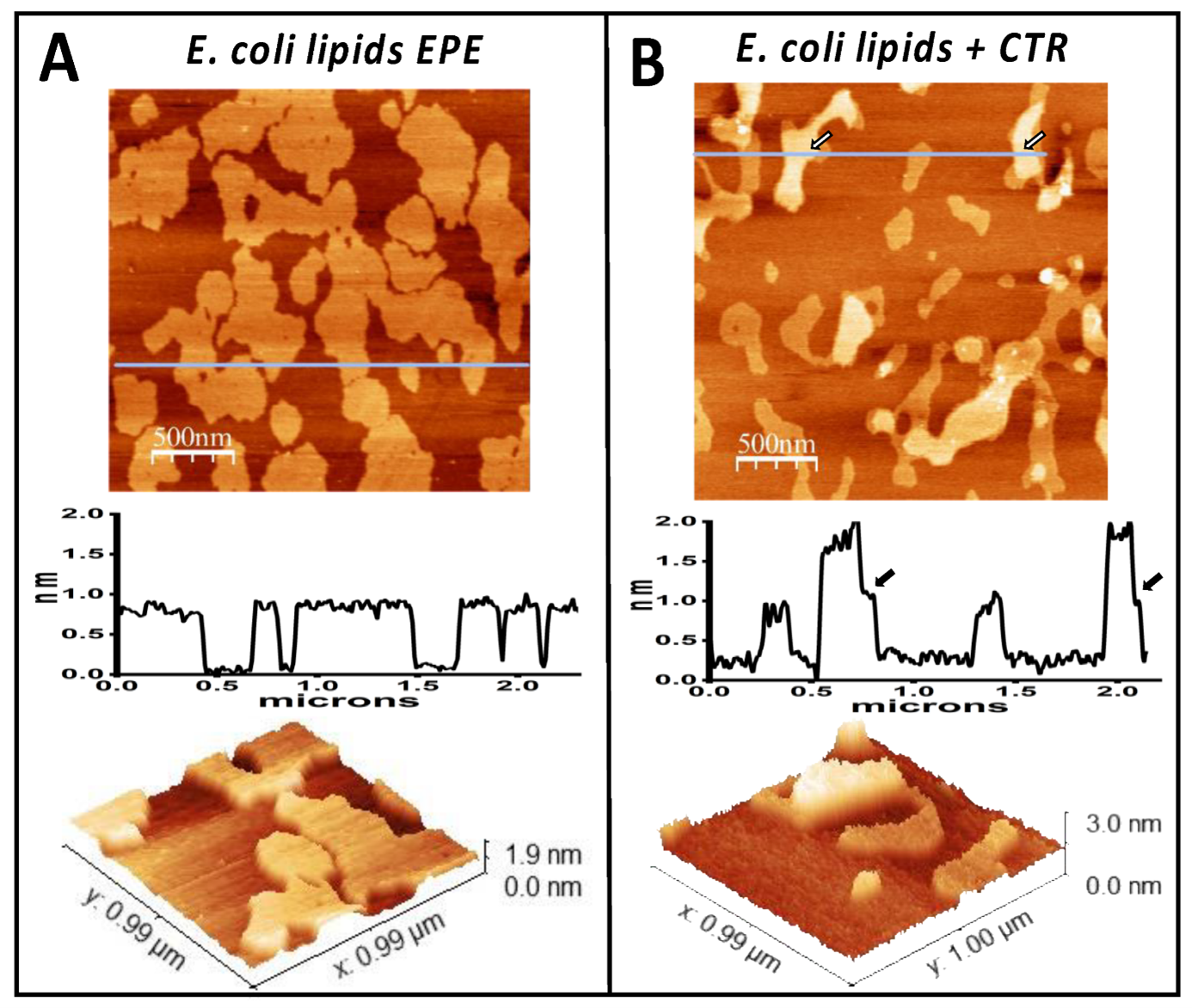
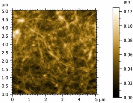
In this article, atomic force microscopy (AFM) confirmed the successful production of microfibrillated cellulose structures with average fibril diameters below 100 nm. The resulting films demonstrated excellent oxygen barrier performance, high stiffness, strong tensile properties, and effective UV-shielding capabilities. Among the investigated materials, films produced from coffee silverskin exhibited particularly promising performance, highlighting the potential of converting agricultural by-products into value-added packaging materials.
To investigate film surface topography, AFM measurements were performed in contact resonance amplitude imaging mode using a NanoWorld Arrow-FMR AFM probe. The AFM probe features a rectangular cantilever with a triangular free end and a tetrahedral tip with a typical radius of curvature of approximately 10 nm. With a spring constant of 2.8 N/m and a resonance frequency of 75 kHz, the Arrow-FMR AFM probe enabled detailed nanoscale characterization of film morphology and surface roughness.
The study demonstrates how AFM analysis using a NanoWorld AFM probe contributes to understanding the relationship between cellulose microstructure and the functional performance of sustainable packaging materials. The results further support the development of renewable, high-performance cellulosic thin films derived from waste biomass sources.
Full citation:
Bellesia, T.; Carullo, D.; Fachin, A.; Soltanzadeh, M.; Ghaani, M.; Ascrizzi, G. I.; Piazza, L.; Farris, S.
Microfibrillated cellulose films from agri-food wastes and plant residues for food packaging applications – A comparative investigation.
Food Packaging and Shelf Life 2026, 54, 101728.
https://doi.org/10.1016/j.fpsl.2026.101728
Attribution 4.0 International
https://creativecommons.org/licenses/by/4.0/