Gastric cancer has a high rate of recurrence, and as such, immunotherapy strategies are being investigated as a potential therapeutic strategy. *
Although the involvement of immune checkpoints in immunotherapy is well studied, biomechanical cues, such as target cell stiffness, have not yet been subject to the same level of investigation. *
Changes in the cholesterol content of the cell membrane directly influence tumor cell stiffness. *
In the article “Cholesterol-regulated cellular stiffness may enhance evasion of NK cell-mediated cytotoxicity in gastric cancer stem cells” Lijuan Zhu and Hongjin Wang investigate the effect of cholesterol on NK cell-mediated killing of gastric cancer stem-like cells. *
They report that surviving tumor cells with stem-like properties elevated cholesterol metabolism to evade NK cell cytotoxicity. *
Inhibition of cholesterol metabolism enhances NK cell-mediated killing of gastric cancer stem-like cells, highlighting a potential avenue for improving immunotherapy efficacy. *
This study suggests a possible effect of cancer cell stiffness on immune evasion and offers insights into enhancing immunotherapeutic strategies against tumors. *
Measurement of cell stiffness by AFM:
A customized commercially available atomic force microscope (AFM) and NanoWorld Pyrex-NitridePNP-TR AFM probes were used for the measurement of cell stiffness by AFM. *
Atomic force microscopy cell stiffness was measured according to standard methodology. *
AFM force curves were captured with a customized AFM placed atop an inverted optical microscope that had a heating stage for live-cell imaging and a ×20 objective. Using an XY stage, the materials were moved until the desired cell, which could be observed under an optical microscope, was positioned beneath the AFM tip. Using a NanoWorld PNP-TR-B AFM cantilever (NanoWorld), the force curves on the cell were collected at a rate of ~ 5 μm·s−1 in the relative trigger mode (15 nm trigger threshold). *
By utilizing a thermal tuning and the deflection sensitivity of 170 nm·V−1, the AFM cantilever spring constant was determined to be 0.08 N·m−1. *
Single cells were measured both before and after treatment at 37 °C. The force curves were processed using the AFM’s analysis software, which also computed the Young’s modulus of the sample. This was accomplished by fitting the approach curve to an indentation of less than 500 nm (to account for stiffness) and assuming a cortical Poisson’s ratio of 0.3.*
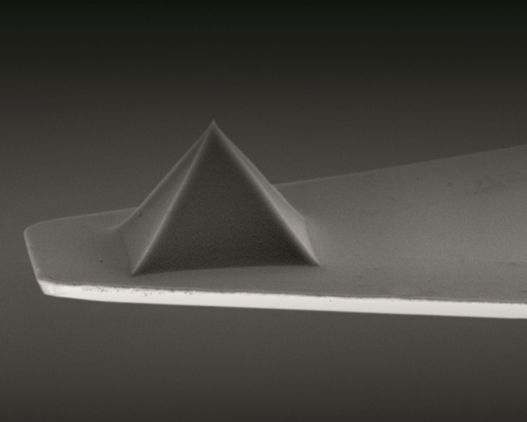
NanoWorld Pyrex-Nitride AFM probe series – AFM tip and AFM cantilever made of silicon nitride
*Lijuan Zhu and Hongjin Wang Cholesterol-regulated cellular stiffness may enhance evasion of NK cell-mediated cytotoxicity in gastric cancer stem cells
FEBS Open Bio, Volume 14, Issue 5, May 2024, Pages 855-866
DOI: https://doi.org/10.1002/2211-5463.13793
Open Access The article “Cholesterol-regulated cellular stiffness may enhance evasion of NK cell-mediated cytotoxicity in gastric cancer stem cells” by Lijuan Zhu and Hongjin Wang is licensed under a Creative Commons Attribution 4.0 International License, which permits use, sharing, adaptation, distribution and reproduction in any medium or format, as long as you give appropriate credit to the original author(s) and the source, provide a link to the Creative Commons license, and indicate if changes were made. The images or other third party material in this article are included in the article’s Creative Commons license, unless indicated otherwise in a credit line to the material. If material is not included in the article’s Creative Commons license and your intended use is not permitted by statutory regulation or exceeds the permitted use, you will need to obtain permission directly from the copyright holder. To view a copy of this license, visit http://creativecommons.org/licenses/by/4.0/.
Airflow limitation in obstructive airway disease is characterized by narrowing of the airway lumen from excessive contraction of airway smooth muscle (ASM) and remodeling of the airway wall which includes changes in the extracellular matrix (ECM) of the ASM layer.*
Previous studies on human airway smooth muscle cells ( hASMC ) have independently assessed the influence of extracellular matrix (ECM) proteins on substrates of supra-physiological stiffnesses, such as tissue culture plastic or glass.*
While the influence of discrete substrate stiffness on hASMC behavior has been examined, manipulation of both substrate stiffness and ECM proteins simultaneously (as expected in disease) has not been extensively modeled in vitro.*
In the article “Stiffness Mediated-Mechanosensation of Airway Smooth Muscle Cells on Linear Stiffness Gradient Hydrogels” Yong Hwee Tan, Kimberley C. W. Wang, Ian L. Chin, Rowan W. Sanderson, Jiayue Li, Brendan F. Kennedy, Peter B. Noble and Yu Suk Choi highlight the interplay and complexities between stiffness and ECM protein type on hASMC mechanosensation, relevant to airway remodelling in obstructive airway diseases.*
The authors first determined a physiological range of ASM layer stiffness using a porcine airway and used these empirical recordings to inform the fabrication of a linear stiffness gradient platform coated with different ECM proteins.*
Using this linear stiffness gradient platform, Yong Hwee Tan et al. profiled hASMC morphology, contractile function with alpha-smooth muscle actin (αSMA) and mechanisms of mechanosensation, specifically with nuclear translocalization of Yes-associated protein (YAP) and lamin-A expression.*
Yong Hwee Tan et al.’s assessment of hASMC mechanosensation utilized an innovative hydrogel platform delivering a linear stiffness gradient to understand stiffness-mediated cell behavior with an ECM substrate for cellular adhesion. *
The employment of a stiffness gradient that was designed after empirical measurements performed on ex vivo ASM tissue, enabled the presentation of physiologically relevant stiffnesses to study hASMC behavior.*
Using this platform, the authors of the article found that hASMC mechanosense underlying mechanical cues more than the types of proteins they are anchored to by screening hASMC morphology, contractile phenotype, and mechanomarker expression, with a few exceptions.*
While the authors acknowledge that the findings from their study were done using cells from only one donor they still think that their study provides a proof of concept for the relevance of hASMC mechanosensation to ECM stiffness, and is another step in the right direction for understanding the pathophysiological impact of airway remodeling in obstructive diseases and exploring potential avenues for improving therapy through greater fidelity of in vitro platforms that include key concepts of mechanosensation. *
Yong Hwee Tan et al. wanted to use the same method which is used to assess hydrogel stiffness, namely atomic force microscopy (AFM), to measure ASM stiffness.*
However, nanoscale measurements of ASM strips by AFM proved to be difficult due to an uneven tissue surface after de-epithelialization (Figure S1C, Supporting Information of the cited article), resulting in false force triggering.
To validate the translation of stiffness values measured from macroscale compression (ASM strips) to nanoscale indentation (AFM on hydrogels), Yong Hwee Tan et al. fabricated additional hydrogels of four different stiffnesses using well-characterized polyacrylamide and compared the stiffness of hydrogels measured by uniaxial compression tester and atomic force microscopy (Figure S2A, Supporting Information of the cited article).
The nanoscale stiffness of hydrogels was assessed using an atomic force microscope (AFM) with NanoWorld triangular Pyrex-NitridePNP-TR AFM probes (the longer AFM cantilever beam – CB 2 – with 200 µm length was used).
These AFM tips probed hydrogels immersed in 1 × PBS with 2 nN, an approach velocity of 2 µm s−1 and a retraction velocity of 10 µm s−1.
Young’s modulus was determined from linear portions of contact-generated force curves using a custom-written code in Igor Pro.
All probe indentations were made in triplicate and averaged for a stiffness measurement in kilopascals (kPa).
An example force curve is shown in Figure S2B, Supporting Information of the cited article. Validation of a linear stiffness gradient was achieved with eight indentations on the hydrogel, 2 mm away from both edges of the hydrogel and at 1 mm intervals along the stiffness gradient axis. Measurements were plotted against displacement from the hydrogel edge (soft to stiff) (Figure 2B of the cited article).
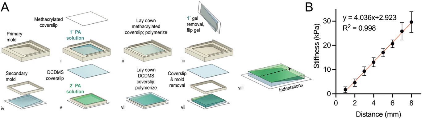
Figure 2 from Yong Hwee Tan et al. 2024 “Stiffness Mediated-Mechanosensation of Airway Smooth Muscle Cells on Linear Stiffness Gradient Hydrogels”: Linear stiffness gradient hydrogel fabrication. A) A schematic of a two-step polymerization process. i) 120 µL of mixed polyacrylamide (PA) solution (% acrylamide + % bis-acrylamide) was added to the primary mold and left to polymerize under ii) a methacrylated coverslip for 20 min. iii) Wedge-shaped 1° gel was removed and flipped for placement of a iv) secondary mold before v) addition of a second 120 µL PA solution and polymerized under a vi) dichlorodimethylsilane-coated coverslip for 20 min. vii) Removal of coverslip and mold completes the fabrication of bi-layered stiffness gradient hydrogel. viii) the dotted arrow indicating the direction of gradient and atomic force microscopy (AFM) measurement. B) Young’s moduli gradient measured by AFM. Twelve hydrogels were selected (one gel per batch) and assessed for stiffness, yielding a gradient of 4.0 kPa mm−1, with a range of 1.7 ± 1.2 to 29.6 ± 4.3 kPa (R2 = 0.998, n = 8). Data are presented as mean ± SD.
Figure S2 from Yong Hwee Tan et al. 2024 “Stiffness Mediated-Mechanosensation of Airway Smooth Muscle Cells on Linear Stiffness Gradient Hydrogels”: (A) Correlation of Young’s modulus from macroscale stiffness (UCT) assessment with nanoindentation (AFM), was conducted using cylindrical PA hydrogels of different Acrylamide %/Bis-acrylamide % derived from Tse and Engler [47] 10 %/0.06 %, 10 %/0.1 %, 10 %/0.15 % and 10 %/0.3 % (Linear regression, P < 0.0001, R2 = 0.9288, n = 4). Data are presented as mean SEM. (B) An example force curve from atomic force microscopy with an approach velocity of 2 μm/s, until a 2 nN trigger force was registered, and retraction of indenter at 10 μm/s.*Yong Hwee Tan, Kimberley C. W. Wang, Ian L. Chin, Rowan W. Sanderson, Jiayue Li, Brendan F. Kennedy, Peter B. Noble and Yu Suk Choi Stiffness Mediated-Mechanosensation of Airway Smooth Muscle Cells on Linear Stiffness Gradient Hydrogels
Advanced Healthcare Materials 2024, 2304254
DOI: https://doi.org/10.1002/adhm.202304254
The article “Stiffness Mediated-Mechanosensation of Airway Smooth Muscle Cells on Linear Stiffness Gradient Hydrogels” by Yong Hwee Tan, Kimberley C. W. Wang, Ian L. Chin, Rowan W. Sanderson, Jiayue Li, Brendan F. Kennedy, Peter B. Noble and Yu Suk Choi is licensed under a Creative Commons Attribution 4.0 International License, which permits use, sharing, adaptation, distribution and reproduction in any medium or format, as long as you give appropriate credit to the original author(s) and the source, provide a link to the Creative Commons license, and indicate if changes were made. The images or other third-party material in this article are included in the article’s Creative Commons license, unless indicated otherwise in a credit line to the material. If material is not included in the article’s Creative Commons license and your intended use is not permitted by statutory regulation or exceeds the permitted use, you will need to obtain permission directly from the copyright holder. To view a copy of this license, visit https://creativecommons.org/licenses/by/4.0/.
The plasticity and growth of plant cell walls (CWs) is still not sufficiently understood on its molecular level. *
Atomic Force Microscopy (AFM) has been shown to be a powerful tool to measure the stiffness of plant tissues. *
In the article “Correlation between plant cell wall stiffening and root extension arrest phenotype in the combined abiotic stress of Fe and Al” Harinderbir Kaur, Jean-Marie Teulon, Christian Godon, Thierry Desnos, Shu-wen W. Chen and Jean-Luc Pellequer describe the use of atomic force microscopy (AFM) to observe elastic responses of the root transition zone of 4-day-old Arabidopsis thaliana wild-type and almt1-mutant seedlings grown under Fe or Al stresses. *
In order to evaluate the relationship between root extension and root cell wall elasticity, the authors used Atomic Force Microscopy to perform vertical indentations on surfaces of living plant roots. *
NanoWorld Pyrex-Nitride silicon-nitride PNP-TR AFM probes with triangular AFM cantilevers were used for the nanoindentation experiments with atomic force microscopy. (PNP-TR AFM cantilever beam 2 (CB2) with a typical force constant of 0.08 N/m and a typical resonant frequency of 17 kHz, typical AFM tip radius 10 nm, macroscopic half cone angles 35°). *
Force-distance (F-D) curves were measured using the Atomic Force Microscope and the PNP-TR AFM tips. *
Because of the heterogeneity of seedling CW surfaces, Harinderbir Kaur et al. used the recently developed trimechanics-3PCS framework for interpreting force-distance curves. The trimechanics-3PCS framework allows the extraction of both stiffness and elasticity along the depth of indentation and permits the investigation of the variation of stiffness with varied depth for biomaterials of heterogeneous elasticity responding to an external force. *
A glass slide with a glued seedling (see Figure 1 cited below) was positioned under the AFM cantilever with the help of an AFM optical camera. Due to the large motorized sample stage of the AFM, the glass slide was adjusted in such a way that the AFM cantilever could be positioned perpendicularly at the longitudinal middle of the glued root. The target working area, the transition zone, was 500 µm away from the root apex, almost twice the length of PNP-TR AFM cantilever. *
As shown in the article the presence of single metal species Fe2+ or Al3+ at 10 μM exerts no noticeable effect on the root growth compared with the control conditions. On the contrary, a mix of both the metal ions produced a strong root-extension arrest concomitant with significant increase of CW stiffness. *
Raising the concentration of either Fe2+or Al3+ to 20 μM, no root-extension arrest was observed; nevertheless, an increase in root stiffness occurred. In the presence of both the metal ions at 10 μM, root-extension arrest was not observed in the almt1 mutant, which substantially abolishes the ability to exude malate. The authors’ results indicate that the combination of Fe2+and Al3+ with exuded malate is crucial for both CW stiffening and root-extension arrest. *
It is shown that the elasticity of plant CW is sensitive and can be used to assess abiotic stresses on plant growth and stiffening. *
However, stiffness increase induced by single Fe2+ or Al3+ is not sufficient for arresting root growth in the described experimental conditions and unexpectedly, the stiffening and the phenotype of seedling roots such as REA are not directly correlated. *
Figure 1 from Harinderbir Kaur et al. 2024 “Correlation between plant cell wall stiffening and root extension arrest phenotype in the combined abiotic stress of Fe and Al”: Principle of nanomechanical measurement of seedling roots with atomic force microscopy. A seedling root (R) is deposited on a microscope slide using silicon glue (N, for Nusil). A fastening band of silicon is seen near the tip of the root (T). The thickness of the fastening band must be thin enough to avoid hindering the AFM support (S), but thick enough to withstand the bending of the root tip. The root is placed under the AFM cantilever (C) as observed by the AFM optical camera. The triangular shaped cantilever (200 µm long) was placed 500 µm away from the root tip in the transition zone where nanoindentation measurements proceeded (as shown). The seedling root and the AFM cantilever are placed within a liquid environment (growth solution, see Supplementary file of the cited article). AFM, atomic force microscopy.
*Harinderbir Kaur, Jean‐Marie Teulon, Christian Godon, Thierry Desnos, Shu‐wen W. Chen and Jean‐Luc Pellequer Correlation between plant cell wall stiffening and root extension arrest phenotype in the combined abiotic stress of Fe and Al
Plant, Cell & Environment 2024; 47:574–584
DOI: https://doi.org/10.1111/pce.14744
The article “Correlation between plant cell wall stiffening and root extension arrest phenotype in the combined abiotic stress of Fe and Al” by Harinderbir Kaur, Jean‐Marie Teulon, Christian Godon, Thierry Desnos, Shu‐wen W. Chen and Jean‐Luc Pellequer is licensed under a Creative Commons Attribution 4.0 International License, which permits use, sharing, adaptation, distribution and reproduction in any medium or format, as long as you give appropriate credit to the original author(s) and the source, provide a link to the Creative Commons license, and indicate if changes were made. The images or other third-party material in this article are included in the article’s Creative Commons license, unless indicated otherwise in a credit line to the material. If material is not included in the article’s Creative Commons license and your intended use is not permitted by statutory regulation or exceeds the permitted use, you will need to obtain permission directly from the copyright holder. To view a copy of this license, visit https://creativecommons.org/licenses/by/4.0/.
![Figure S2 from Yong Hwee Tan et al. 2024 “Stiffness Mediated-Mechanosensation of Airway Smooth Muscle Cells on Linear Stiffness Gradient Hydrogels”:(A) Correlation of Young’s modulus from macroscale stiffness (UCT) assessment with nanoindentation (AFM), was conducted using cylindrical PA hydrogels of different Acrylamide %/Bis-acrylamide % derived from Tse and Engler [47] 10 %/0.06 %, 10 %/0.1 %, 10 %/0.15 % and 10 %/0.3 % (Linear regression, P < 0.0001, R2 = 0.9288, n = 4). Data are presented as mean SEM. (B) An example force curve from atomic force microscopy with an approach velocity of 2 μm/s, until a 2 nN trigger force was registered, and retraction of indenter at 10 μm/s. NanoWorld triangular Pyrex-Nitride PNP-TR AFM probes were used to assess the stiffness of the hydrogels with atomic force microscopy.](https://www.nanoworld.com/blog/wp-content/uploads/2024/07/Fig-S2-from-Young-Hwee-Tan-2024-Stiffness-Mediated-Mechanosensation-of-Airway-Smooth-Muscle-Cells-on-Linear-Stiffness-Gradient-Hydrogels-NanoWorld-PNP-TR-AFM-probe.jpg)
