A beginner’s guide to the Characterization of Hydrogel Microarchitecture for Cellular Applications

Hydrogel materials show a number of properties which make them interesting candidates to be utilized to mimic the extracellular matrix (ECM). Therefore, these materials are attractive for use in biological applications such as tissue engineering, cell culture 3D bioprinting and more.

Are you planning to use hydrogels for the first time in your research?

Then have a look at the insightful article “A beginner’s guide to the Characterization of Hydrogel Microarchitecture for Cellular Applications” by Francisco Drusso Martinez-Garcia, Tony Fischer, Alexander Hayn, Claudia Tanja Mierke, Janette Kay Burgess and Martin Conrad Harmsen.

In their article the authors describe and evaluate the different technologies that are most commonly used to assess hydrogel microarchitecture.

Francisco Drusso Martinez-Garcia et al. explain the working principle of the various methods and also discuss the merits and limitations of each of them in view of their usefulness for the characterization of hydrogels.

They introduce and explore the pros and cons of the following methods: Scanning Electron Microscopy (SEM), Cryogenic Scanning Electron Microscopy (Cryo-SEM), Environmental Scanning Electron Microscopy (ESEM), Micro-Computed Tomography (µ-CT), Confocal Laser Scanning Microscopy (CLSM), Second Harmonic Generation and Atomic Force Microscopy (AFM).*

Atomic force microscopy (AFM) can be used to investigate the hydrogel surface topology as well as a hydrogel’s mechanical properties. The latter can be achieved through mathematical modelling of force-distance curves.

When using the AFM to characterize the elasticity of a hydrogel sample it is essential to take the stiffness of the investigated material into account when choosing what kind of AFM probe to use for these experiments.

If an AFM cantilever used for probing a soft sample is too stiff (if the force constant/spring constant is too high) this might result in a poor signal-to-noise ratio.

If a soft AFM probe (an AFM probe with an AFM cantilever with a low force constant) is chosen to investigate a soft material this should lead to a better signal-to-noise ratio. On the other hand, if an AFM cantilever is too soft (if the force constant is too low) then it might not be stiff enough to indent the investigated material.

Another critical factor is the shape and the size of the AFM tip.

Spheroidal AFM probes might stick to the material, resulting in artefacts, disrupted force–distance curves, or even damaged AFM cantilevers. If the AFM tip is much smaller than the pore size of the hydrogel, it might get stuck in the fibrous network microarchitecture.

On the other hand, if the spherical AFM tip, e.g. as in colloidal AFM probes (a sphere glued to end of a tipless AFM cantilever), is too large, the weight of the sphere can have a negative influence on the spring characteristics of the AFM cantilever.

All these factors and more as described in the cited article have to be carefully weighed before deciding on the settings of the atomic force microscope and choosing an AFM probe for the investigation of a specific hydrogel.

NanoWorld tipless ArrowTL2 cantilever arrays with polystyrene beads glued to them were used by the authors of this beginner’s guide to achieve the AFM data presented in the article.*
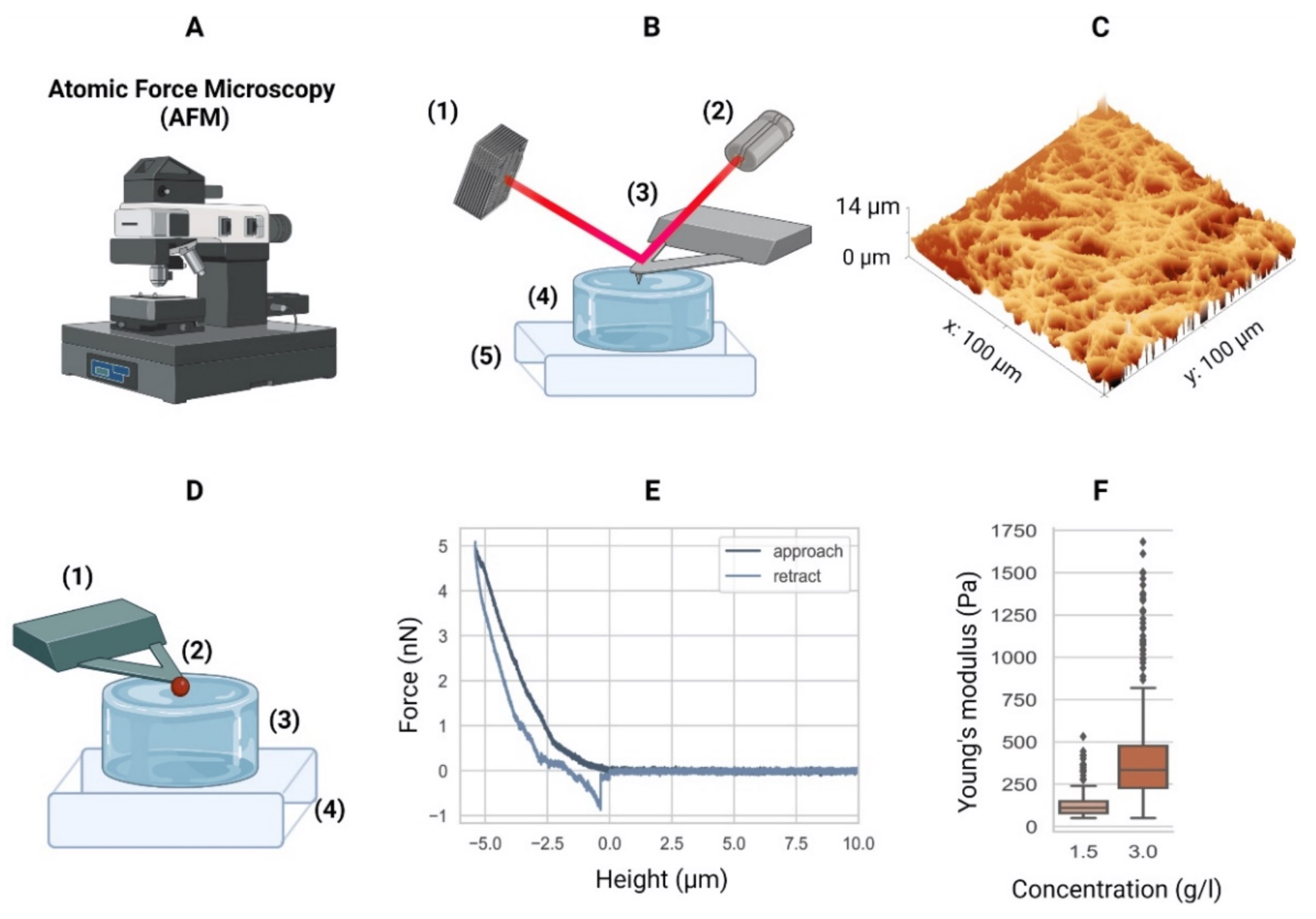
Figure 6. from Francisco Drusso Martinez-Garcia et al. 2022: Atomic force microscopy. (A) Equipment. (B) Schematic of an AFM setup with a four-quadrant photodiode (1), in which the four-quadrant photodiode (1) receives a laser (2) reflected from a cantilever (3), in this case positioned over a hydrogel (4) mounted in a piezo stage (5). For example, the height differences in a sample (4) are measured by adjusting the stage using piezo elements (5) to counter the cantilever bending on a nanometer scale. (C) The AFM can then generate a surface heightmap of the hydrogels such as a GelMA hydrogel (shown). AFM can also be used to determine the mechanical properties of hydrogels. (D) Schematic of the AFM technique to determine the elastic moduli of hydrogels with a tipless cantilever (1), spheroidal probe (2, red), hydrogel (3), and stiff substrate (4). As the cantilever represents a spring with a known spring constant, the cantilever bending due to elastic counterforces exerted by the soft material is correlated with the piezo stage height (4). (E) The so-called force–distance curves are recorded. Data from a collagen type-I hydrogel (3.0 g/L) are shown. (F) Young’s moduli of a 1.5 g/L and 3.0 g/L collagen type-I hydrogel. Outliers indicated by ◆. AFM equipment detailed in Appendix A of the cited article. NanoWorld tipless ArrowTL2 cantilever arrays with polystyrene beads glued to them were used by the authors of this beginner’s guide to achieve the AFM data presented in the article.
Figure 6. from Francisco Drusso Martinez-Garcia et al. 2022:
Atomic force microscopy. (A) Equipment. (B) Schematic of an AFM setup with a four-quadrant photodiode (1), in which the four-quadrant photodiode (1) receives a laser (2) reflected from a cantilever (3), in this case positioned over a hydrogel (4) mounted in a piezo stage (5). For example, the height differences in a sample (4) are measured by adjusting the stage using piezo elements (5) to counter the cantilever bending on a nanometer scale. (C) The AFM can then generate a surface heightmap of the hydrogels such as a GelMA hydrogel (shown). AFM can also be used to determine the mechanical properties of hydrogels. (D) Schematic of the AFM technique to determine the elastic moduli of hydrogels with a tipless cantilever (1), spheroidal probe (2, red), hydrogel (3), and stiff substrate (4). As the cantilever represents a spring with a known spring constant, the cantilever bending due to elastic counterforces exerted by the soft material is correlated with the piezo stage height (4). (E) The so-called force–distance curves are recorded. Data from a collagen type-I hydrogel (3.0 g/L) are shown. (F) Young’s moduli of a 1.5 g/L and 3.0 g/L collagen type-I hydrogel. Outliers indicated by ◆. AFM equipment detailed in Appendix A of the cited article.


NanoWorld tipless Arrow-TL2 AFM probe array with two tipless AFM cantilevers
NanoWorld® Arrow™ TL2 AFM probes are tipless AFM cantilevers for special applications. They can for example be used for attaching spheres and other objects to the free end of the AFM cantilever, or for functionalizing and sensing applications.
The Arrow™ TL2 probes are optionally available with a sample facing side gold coating (Arrow™ TL2Au).

*Francisco Drusso Martinez-Garcia, Tony Fischer, Alexander Hayn, Claudia Tanja Mierke, Janette Kay Burgess and Martin Conrad Harmsen
A Beginner’s Guide to the Characterization of Hydrogel Microarchitecture for Cellular Applications
Gels 2022, 8(9), 535
DOI: https://doi.org/10.3390/gels8090535

The article “A Beginner’s Guide to the Characterization of Hydrogel Microarchitecture for Cellular Applications” by Francisco Drusso Martinez-Garcia, Tony Fischer, Alexander Hayn, Claudia Tanja Mierke, Janette Kay Burgess and Martin Conrad Harmsen is licensed under a Creative Commons Attribution 4.0 International License, which permits use, sharing, adaptation, distribution and reproduction in any medium or format, as long as you give appropriate credit to the original author(s) and the source, provide a link to the Creative Commons license, and indicate if changes were made. The images or other third-party material in this article are included in the article’s Creative Commons license, unless indicated otherwise in a credit line to the material. If material is not included in the article’s Creative Commons license and your intended use is not permitted by statutory regulation or exceeds the permitted use, you will need to obtain permission directly from the copyright holder. To view a copy of this license, visit https://creativecommons.org/licenses/by/4.0/.

High-speed atomic force microscopy highlights new molecular mechanism of daptomycin action

The current pandemic is not the only health threat worldwide. Another worry is the increasing antibiotic resistance which increases the fear to run out of effective antibiotics.

This is one of the reasons why antimicrobial peptides (AMPs) are gaining more and more interest.

The lipopeptide Daptomycin ( DAP ) has been therapeutically used as a last resort antibiotic against multidrug-resistant enterococci and staphylococci in the past. Unfortunately, some strains have become resistant to Dap in recent years. There still is a knowledge-gap on the details of Dap activity. It is therefore important to understand the structure-activity relationships of AMPs on membranes in order to develop more antibiotics of this type as a countermeasure to the spread of resistance.*

High Speed Atomic Force Microscopy ( HS-AFM ) makes it possible to observe dynamic biological processes on a molecular level.

In the article “High-speed atomic force microscopy highlights new molecular mechanism of daptomycin action” Francesca Zuttion, Adai Colom, Stefan Matile, Denes Farago, Frédérique Pompeo, Janos Kokavecz, Anne Galinier, James Sturgis and Ignacio Casuso describe how, by using the possibilities offered by high speed atomic force microscopy, they were able to confirm some up until now hypothetical models and additionally detected some previously unknown molecular mechanisms. *

The HS-AFM imaging made it possible for the authors to observe the development of the dynamics of interaction at the molecular-level over several hours. *

They investigated the lipopeptide Daptomycin under infection-like conditions and could confirm Dap oligomerization and the existence of half pores. *

They also mimicked bacterial resistance conditions by increasing the CL-content in the membrane. *

By correlating the results of other research techniques such as FRET, SANS, NMR, CD or electrophysiology techniques with the results they achieved with high speed atomic force microscopy F. Zuttion et al. were able to confirm several, previously, hypothetical models, and detect several unknown molecular mechanisms. *

It is to be hoped that the possibilities offered by HS-AFM imaging will stimulate new models and insight on the structure-activity relationship of membrane-interacting molecules and also open up the possiblity to increase the throughput of screening of molecular candidates considerably. *

NanoWorld USC ( Ultra-Short AFM Cantilevers) of the USC-F1.2-k0.15 type, which are specially designed for the use in high speed atomic force microscopy, were used for the HS-AFM imaging described in the article cited below. These AFM probes have a typical resonance frequency of 1200 kHz and have a wear resistant AFM tip made from high density carbon.

Figure 4 Sub-MIC Dap on POPG at 37 °C. Tens of minutes from “High-speed atomic force microscopy highlights new molecular mechanism of daptomycin action” by Francesca Zuttion et al. NanoWorld Ultra-Short AFM Cantilevers USC-F1.2-k0.15 AFM probes for HS-AFM imaging were used.
Figure 4 Sub-MIC Dap on POPG at 37 °C. Tens of minutes from “High-speed atomic force microscopy highlights new molecular mechanism of daptomycin action” by Francesca Zuttion et al.
Intermediate stages a A new structure appeared: dimples, zones of thinner membrane thickness, whose diameter was in the range 7 ± 2 nm. Most dimples diffuse, but some remained static (colour scale: 3 nm). Movie details: frame rate 97 ms; zoom of a full image of 150 nm × 90 nm and 256 × 160 pixels. b The dimple diffusion consisted of swinging trajectories, implying membrane-mediated dimple-dimple attraction (colour scale: 3 nm). b, right, Energy profile of the interaction of the dimples obtained derived from 120 centre-to-centre distance measurements that contains as the oligomers two energy minima. Movie details: frame rate 83 ms; full image of 150 nm × 150 nm and 256 × 256 pixels. c In some membrane zones, clusters of dimples, reminiscent of cubic phases, developed (colour scale: 4 nm). Movie details: frame rate 74 ms; full image of 90 nm × 60 nm and 256 × 160 pixels. d The clusters of dimples were moderately dynamical in time, with moderate internal rearrangements (colour scale: 4 nm). Movie details: frame rate 74 ms; full image of 25 nm × 16 nm and 256 × 160 pixels. e The other deformation found was elongated-humps on top of the POPG membrane. e, left, An elongated-hump in the proximity of a cluster of dimples (colour scale: 4 nm). e, right, A close-up and a profile of an elongated-hump. Additional images of elongated-humps on Supplementary Fig. 1. Movie details: frame rate 479 ms; zoom of full image of 250 nm × 200 nm and 300 × 256 pixels. f It was observed that the dimples and the elongated-humps fused and gave yield to pores of toroidal structure where a protruding ring surrounds the pore (colour scale: 4 nm). Movie details: frame rate 74 ms; full image of 40 nm × 40 nm and 256 × 160 pixels.

*Francesca Zuttion, Adai Colom, Stefan Matile, Denes Farago, Frédérique Pompeo, Janos Kokavecz, Anne Galinier, James Sturgis and Ignacio Casuso
High-speed atomic force microscopy highlights new molecular mechanism of daptomycin action
Nature Communications volume 11, Article number: 6312 (2020)
DOI: https://doi.org/10.1038/s41467-020-19710-z

Please follow this external link to read the full article: https://rdcu.be/ciaW2

Open Access : The article “High-speed atomic force microscopy highlights new molecular mechanism of daptomycin action” by Francesca Zuttion, Adai Colom, Stefan Matile, Denes Farago, Frédérique Pompeo, Janos Kokavecz, Anne Galinier, James Sturgis and Ignacio Casuso is licensed under a Creative Commons Attribution 4.0 International License, which permits use, sharing, adaptation, distribution and reproduction in any medium or format, as long as you give appropriate credit to the original author(s) and the source, provide a link to the Creative Commons license, and indicate if changes were made. The images or other third party material in this article are included in the article’s Creative Commons license, unless indicated otherwise in a credit line to the material. If material is not included in the article’s Creative Commons license and your intended use is not permitted by statutory regulation or exceeds the permitted use, you will need to obtain permission directly from the copyright holder. To view a copy of this license, visit https://creativecommons.org/licenses/by/4.0/.

Tissue mechanics and expression of TROP2 in oral squamous cell carcinoma with varying differentiation

Atomic Force Microscopy ( AFM ) can be utilized to determine the mechanical properties of tumor tissues in different kinds of cancers, for example breast cancer, liver cancer and lung cancer.

Oral squamous cell carcinoma (OSCC) is a common subtype of head and neck and other malignant tumors that occurs in increasing numbers. It is therefore important to learn more about the biological factors connected with the early diagnosis and treatment of OSCC. *

The human trophoblast cell surface antigen 2 (TROP2), which is also called tumor-associated calcium signal transduction-2 (TACSTD-2), is a surface glycoprotein encoded by TACSTD. *

Among the various biochemical mechanisms involved in tumorigenesis, the role of β-catenin has been studied extensively. This has shed light on the biological functions of TROP2 and its use as a prognostic biomarker for OSCC. *

TROP2 regulates tumorigenic properties including cancer cell adhesion, invasion, and migration and is overexpressed in many human cancers. Inhibiting TROP2 expression has shown promise in clinical applications. *

In the article “Tissue mechanics and expression of TROP2 in oral squamous cell carcinoma with varying differentiation” Baoping Zhang, Shuting Gao, Ruiping Li, Yiting Li, Rui Cao, Jingyang Cheng, Yumeng Guo, Errui Wang, Ying Huang and Kailiang Zhang investigate the role of TROP2 in OSCC patients using a combination of biophysical approaches including atomic force microscopy. *

The authors demonstrate the tissue morphology and mechanics of OSCC samples during tumor development using NanoWorld Pointprobe® CONTR AFM probes for the Atomic Force Microscopy described in the article and they believe that their findings will help develop TROP2 in accurately diagnosing OSCC in tumors with different grades of differentiation. *

Figure 5 from Baoping Zhang et al. “Tissue mechanics and expression of TROP2 in oral squamous cell carcinoma with varying differentiation”:
Surface morphology of OSCC tissue sections via AFM detection, irregular morphology appeared in the low differentiation
NanoWorld Pointprobe CONTR AFM probes were used for the Atomic Force Microscopy
Figure 5 from Baoping Zhang et al. “Tissue mechanics and expression of TROP2 in oral squamous cell carcinoma with varying differentiation”:
Surface morphology of OSCC tissue sections via AFM detection, irregular morphology appeared in the low differentiation

*Baoping Zhang, Shuting Gao, Ruiping Li, Yiting Li, Rui Cao, Jingyang Cheng, Yumeng Guo, Errui Wang, Ying Huang and Kailiang Zhang
Tissue mechanics and expression of TROP2 in oral squamous cell carcinoma with varying differentiation
BMC Cancer volume 20, Article number: 815 (2020)
DOI: https://doi.org/10.1186/s12885-020-07257-7

Please follow this external link to read the whole article: https://rdcu.be/cfC9G

Open Access : The article “Tissue mechanics and expression of TROP2 in oral squamous cell carcinoma with varying differentiation” by Baoping Zhang, Shuting Gao, Ruiping Li, Yiting Li, Rui Cao, Jingyang Cheng, Yumeng Guo, Errui Wang, Ying Huang and Kailiang Zhang is licensed under a Creative Commons Attribution 4.0 International License, which permits use, sharing, adaptation, distribution and reproduction in any medium or format, as long as you give appropriate credit to the original author(s) and the source, provide a link to the Creative Commons license, and indicate if changes were made. The images or other third party material in this article are included in the article’s Creative Commons license, unless indicated otherwise in a credit line to the material. If material is not included in the article’s Creative Commons license and your intended use is not permitted by statutory regulation or exceeds the permitted use, you will need to obtain permission directly from the copyright holder. To view a copy of this license, visit https://creativecommons.org/licenses/by/4.0/.