In the article «Influence of Ultraviolet Radiation Exposure Time on Styrene-Ethylene-Butadiene-Styrene (SEBS) Copolymer» Daniel Garcia-Garcia , José Enrique Crespo-Amorós, Francisco Parres and María Dolores Samper describe how they studied the effect of ultraviolet radiation on styrene-ethylene-butadiene-styrene (SEBS) at different exposure times in order to obtain a better understanding of the mechanism of ageing.*
The results obtained for the SEBS, in relation to the duration of exposure, showed superficial changes that cause a decrease in the surface energy (s) and, therefore, a decrease in surface roughness. This led to a reduction in mechanical performance, decreasing the tensile strength by about 50% for exposure times of around 200 hours.*
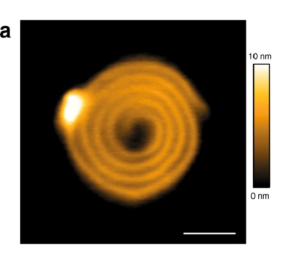
NanoWorld Pointprobe® NCH AFM probes were used in the Atomic force microscopy (AFM) applied to determine the surface topography and roughness of the aged samples.

Figure 4 from «Influence of Ultraviolet Radiation Exposure Time on Styrene-Ethylene-Butadiene-Styrene (SEBS) Copolymer» by Daniel Garcia-Garcia et al.:
Atomic force microscopy (AFM) surface of SEBS: (a) without exposure (T0), (b) UV exposure T2, (c) UV exposure T7 and (d) UV exposure T9.
*Daniel Garcia-Garcia , José Enrique Crespo-Amorós, Francisco Parres and María Dolores Samper
Influence of Ultraviolet Radiation Exposure Time on Styrene-Ethylene-Butadiene-Styrene (SEBS) Copolymer
Polymers 2020, 12, 862
DOI: https://doi.org/10.3390/polym12040862
Please follow this external link to read the full article: https://www.mdpi.com/2073-4360/12/4/862
Open Access : The article “Influence of Ultraviolet Radiation Exposure Time on Styrene-Ethylene-Butadiene-Styrene (SEBS) Copolymer» byDaniel Garcia-Garcia , José Enrique Crespo-Amorós, Francisco Parres and María Dolores Samper is licensed under a Creative Commons Attribution 4.0 International License, which permits use, sharing, adaptation, distribution and reproduction in any medium or format, as long as you give appropriate credit to the original author(s) and the source, provide a link to the Creative Commons license, and indicate if changes were made. The images or other third party material in this article are included in the article’s Creative Commons license, unless indicated otherwise in a credit line to the material. If material is not included in the article’s Creative Commons license and your intended use is not permitted by statutory regulation or exceeds the permitted use, you will need to obtain permission directly from the copyright holder. To view a copy of this license, visit http://creativecommons.org/licenses/by/4.0/.