Ferroelectric polymer thin films crystallize in different phases and microstructures depending on fabrication conditions such as annealing temperature or layer deposition technique.*
In the article “Spherulite-like microstructure observed for spin-cast P(VDF-TrFE) thin films and their ferroelectric characteristics” Davide Disnan, Jonas Hafner, Michael Schneider and Ulrich Schmid demonstrate how the morphology of spin-cast poly(vinylidene fluoride-trifluoroethylene) (P(VDF-TrFE)) thin films changes as a function of annealing temperature from rice-like to spherulite-like microstructure, whereas the latter morphology is closer to the crystallization characteristic of poly(vinylidene-fluoride) (PVDF).*
Thin films of PVDF and P(VDF-TrFE) were analyzed at the nanometre-scale using atomic force microscopy. *
NanoWorld Pyrex-Nitride PNP-TR AFM probes were used for the ferroelectric characterization of the polymer thin films by atomic force microscopy. *
The displacement of the metal-ferroelectric-metal structure in response to the electric field applied was measured at one single point in the centre of the capacitor. For that purpose, the AFM cantilever (NanoWorld PNP-TR with a spring constant of k = 0.32 N/m made of silicon nitride (non-conductive cantilever for avoiding electrical interference caused by ground loops) was used.*
The deflection during the electrical stimulation was calibrated through the measurement of the inverse optical lever sensitivity of the probe. In order to avoid significant indentation effects, the silicon wafer surface surrounding the capacitor structure was used to land the cantilever for the calibration. *
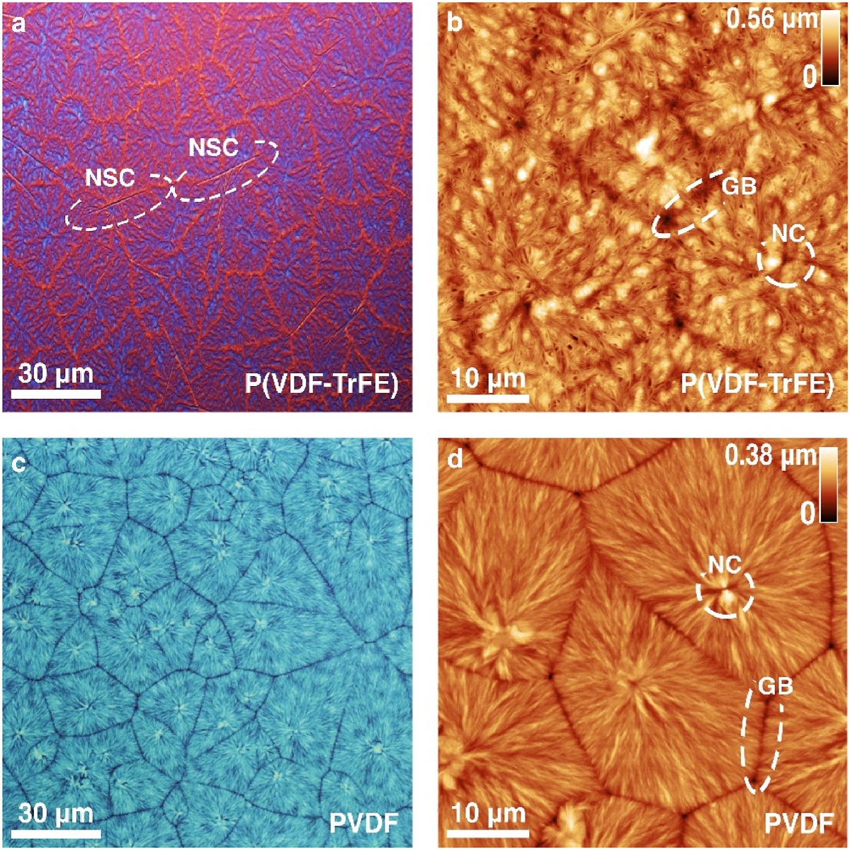
Spherulite-like microstructure of P(VDF-TrFE) and spherulite microstructure of PVDF. a, b Optical micrograph and AFM height image of the spherulite-like microstructure of P(VDF-TrFE). Features like needle-shaped crystals (NSC), nucleation centres (NC) and grain boundaries (GB) are highlighted on the spherulite-like microstructure surface. C ,d Analog for the spherulite microstructure of PVDF.
Please follow the external link to read the full article:
*Davide Disnan, Jonas Hafner, Michael Schneider and Ulrich Schmid
Spherulite-like microstructure observed for spin-cast P(VDF-TrFE) thin films and their ferroelectric characteristics
Polymer, Volume 272, 17 April 2023, 125840
DOI: https://doi.org/10.1016/j.polymer.2023.125840
The article “Spherulite-like microstructure observed for spin-cast P(VDF-TrFE) thin films and their ferroelectric characteristics” by Davide Disnan, Jonas Hafner, Michael Schneider and Ulrich Schmid is licensed under a Creative Commons Attribution 4.0 International License, which permits use, sharing, adaptation, distribution and reproduction in any medium or format, as long as you give appropriate credit to the original author(s) and the source, provide a link to the Creative Commons license, and indicate if changes were made. The images or other third party material in this article are included in the article’s Creative Commons license, unless indicated otherwise in a credit line to the material. If material is not included in the article’s Creative Commons license and your intended use is not permitted by statutory regulation or exceeds the permitted use, you will need to obtain permission directly from the copyright holder. To view a copy of this license, visit https://creativecommons.org/licenses/by/4.0/.