The surface layer (S-layer) of probiotic bacteria plays an important role in their interaction with the host immune system. In this article, Valentina Taverniti , Paolo D’Incecco , Stefano Farris , Peter Riber Jonsen , Helene Skovsted Eld , Juliane Sørensen, Laura Brunelli, Giacomo Mantegazza, Stefania Arioli and Hanne Frøkiær, investigated how the thickness of the S-layer influences the ability of Lactobacillus helveticus MIMLh5 and Lactobacillus acidophilus NCFM to stimulate Th1-related cytokine production in dendritic cells.
The results revealed an inverse correlation between S-layer thickness and the induction of interleukin-12, indicating that thinner S-layers are associated with a stronger immune-stimulating response. These findings provide new insights into the structure–function relationship of bacterial surface layers and their role in probiotic–host interactions.
Atomic force microscopy (AFM) was used for nanomechanical and morphological characterization of bacterial cells. Measurements were performed using a commercially available AFM instrument operated in contact resonance amplitude imaging (CRAI) mode. An Nanoworld Arrow-FMR force modulation AFM probe was used. This silicon AFM probe features a rectangular beam with a triangular free end and a tetrahedral tip (tip radius ~10 nm, tip height 10–15 μm), a spring constant of 2.8 N/m and a resonance frequency of 75 kHz. Images of 10 × 10 μm² and force–distance curves were recorded at multiple locations on the bacterial surface. Nanomechanical properties, including the elastic (Young’s) modulus, were determined by fitting approach curves to the Hertzian model with an indentation depth set to 2 nm.
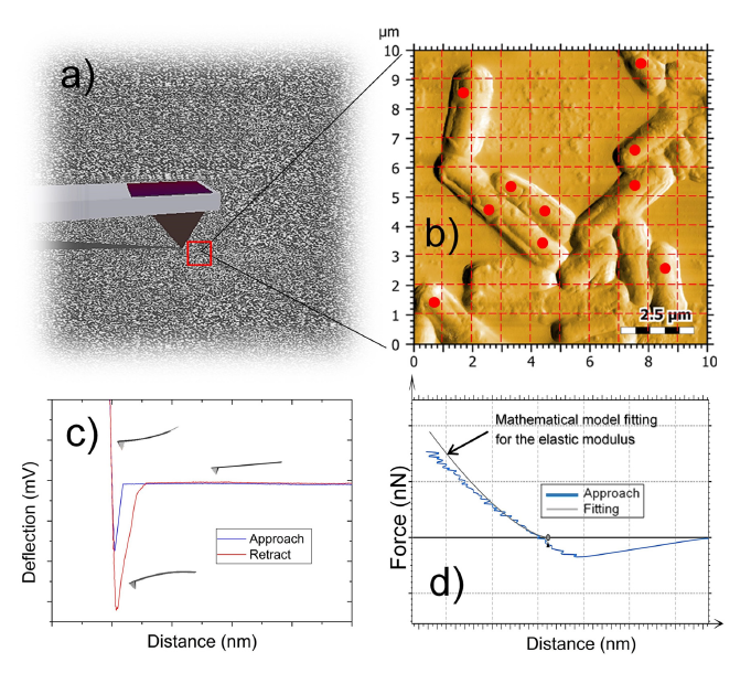
Figure S1: Schematic representation of the 4-step procedure for the AFM analysis of the bacteria surface: scanning of the surface in contact resonance amplitude (CRAI) mode (a); creation of the 10-point map of the nanomechanical test (b); generation of the force-distance curves (c); and fitting procedure for the extrapolation of the elastic modulus (d).
Taverniti, V.; D’Incecco, P.; Farris, S.; Jonsen, P. R.; Eld, H. S.; Sørensen, J.; Brunelli, L.; Mantegazza, G.; Arioli, S.; Mora, D.; Guglielmetti, S.; Frøkiær, H.
The Capacities of the Probiotic Strains L. helveticus MIMLh5 and L. acidophilus NCFM to Induce Th1-Stimulating Cytokines in Dendritic Cells Are Inversely Correlated with the Thickness of Their S-Layers.
Biomolecules 2025, 15(7), 1012.
https://doi.org/10.3390/biom15071012
The article: The Capacities of the Probiotic Strains L. helveticus MIMLh5 and L. acidophilus NCFM to Induce Th1-Stimulating Cytokines in Dendritic Cells Are Inversely Correlated with the Thickness of Their S-Layers, is licensed under a Creative Commons Attribution 4.0 International License, which permits use, sharing, adaptation, distribution and reproduction in any medium or format, as long as you give appropriate credit to the original author(s) and the source, provide a link to the Creative Commons license, and indicate if changes were made. The images or other third party material in this article are included in the article’s Creative Commons license, unless indicated otherwise in a credit line to the material. If material is not included in the article’s Creative Commons license and your intended use is not permitted by statutory regulation or exceeds the permitted use, you will need to obtain permission directly from the copyright holder. To view a copy of this license, visit http://creativecommons.org/licenses/by/4.0/.