Flexible Polyurethane Foams Modified with Novel Coconut Monoglycerides-Based Polyester Polyols

The products of the polyurethane (PU) industry such as foams, coatings and adhesives are numerous and can be found in many areas of everyday life. *

Polyols are an essential component in the production of polyurethane. Nowadays they mostly come from petroleum products. *

In view of potential risk factors such as the running out of fossil fuels, supply chain issues, environmental concerns and economic risks it is important to develop alternatives as substitutes and supplements to the existing petroleum derived polyols. *

Vegetable oils can be used to manufacture biobased polyols and various oils such as linseed oil, rapeseed oil, canola oil, grapeseed oil, corn oil, rice bran oil, palm oil, olive oil, castor oil and soybean oil have already been used to make polyols for different purposes.*

Most of the polyols derived from vegetable oils that are already commercially available are made from soybean and castor oil and are mainly used for rigid PU foam applications. *

So far biobased materials for flexible polyurethane foams (FPUFs) have not been studied as much as their rigid counterpart. This is because, due to their chemical composition, there are limits to how much biobased materials can be used in the flexible foam without having an undesired effect on the foam’s mechanical properties. *

Coconut oil is not often used to manufacture flexible foam because the coconut oil’s high level of saturation makes it less compatible with many common methods of creating polyols, as the widely used polyol-forming processes mostly rely on the unsaturation of vegetable oil for functionalization.

To make coconut monoglycerides (CMG) or other plant-based oils usable for polyol-forming processes they need to fulfil the same structural requirements as the fossil-based products. *

In the article “Flexible Polyurethane Foams Modified with Novel Coconut Monoglycerides-Based Polyester Polyols “ Christine Joy M. Omisol, Blessy Joy M. Aguinid, Gerson Y. Abilay, Dan Michael Asequia, Tomas Ralph Tomon, Karyl Xyrra Sabulbero, Daisy Jane Erjeno, Carlo Kurt Osorio, Shashwa Usop, Roberto Malaluan, Gerard Dumancas, Eleazer P. Resurreccion, Alona Lubguban, Glenn Apostol, Henry Siy, Arnold C. Alguno, and Arnold Lubguban describe how they investigated the potential of coconut monoglycerides (CMG) as a polyol raw material specifically for flexible polyurethane foam (FPUF) applications.*

The authors synthesized high-molecular-weight polyester polyols from coconut monoglycerides (CMG), a coproduct of fatty acid production from coconut oil, via polycondensation at different mass ratios of CMG with 1:5 glycerol:phthalic anhydride.*

The resulting CMG-based polyols were shown to work well in making flexible foam. *

Fourier transform infrared (FTIR) spectroscopy and atomic force microscopy (AFM) were used for the foam characterization. *

The modification of the foam formulation increased the monodentate and bidentate urea groups, shown using Fourier transform infrared (FTIR) spectroscopy, that promoted microphase separation in the foam matrix, confirmed using atomic force microscopy (AFM) and differential scanning calorimetry (DSC). *

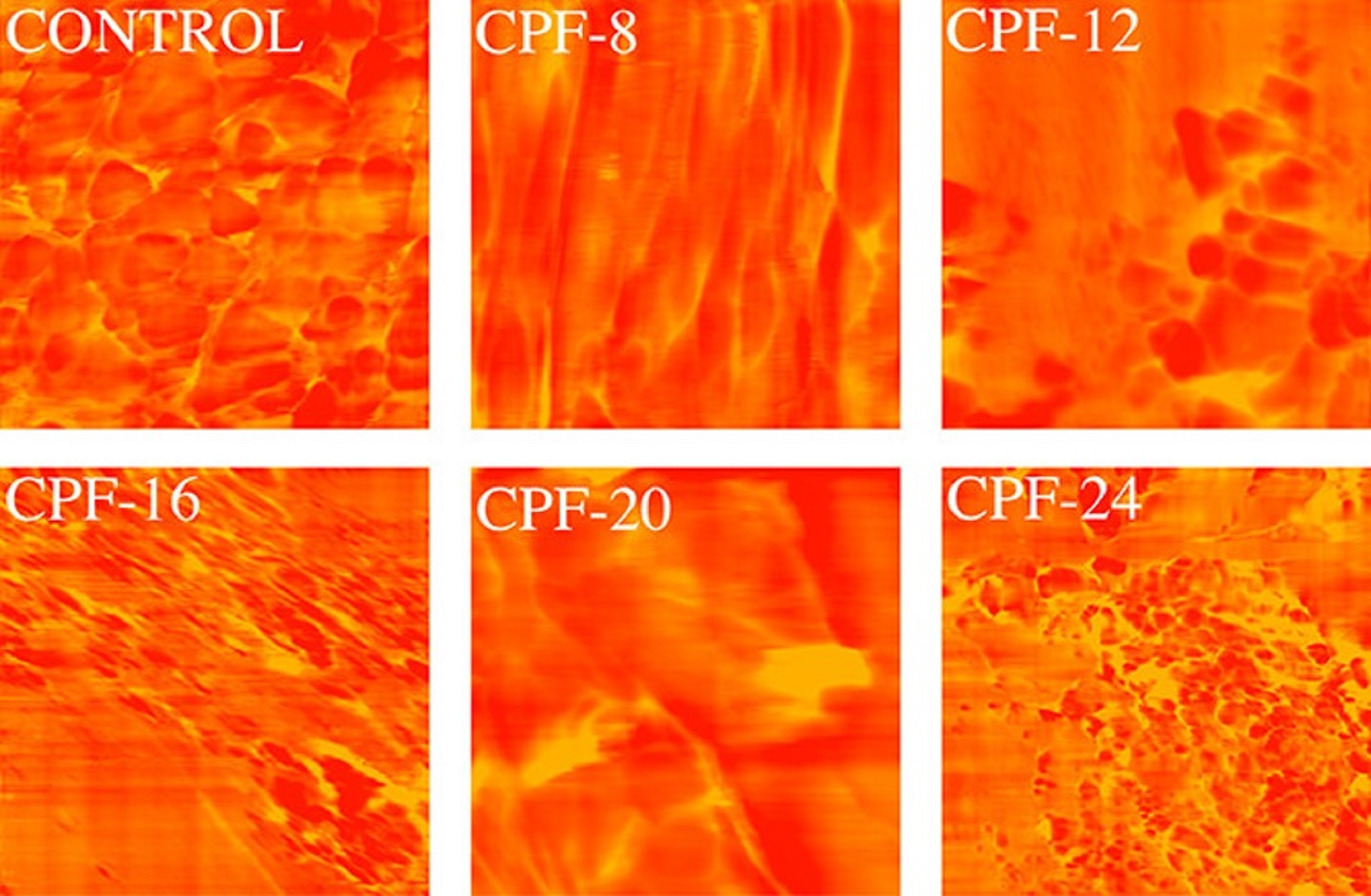
Atomic force microscopy (AFM) was used to evaluate the hard–soft domains phase separation of the foam. *

The atomic force microscope was operated at a scan rate of 1.0 Hz in non-contact mode using NanoWorld Pointprobe® NCHR Silicon AFM probes for standard tapping mode applications. (typical resonance frequency 320 kHz, typical force constant 42 N/m). *

It could be shown that density of the CMGPOL-modified polyurethane foams (CPFs) decreased, while a significant improvement in their tensile and compressive properties was observed. *

The investigations by Christine Joy M. Omisol et al. resulted in a new sustainable polyol raw material that can be used to modify petroleum-based foam and produce flexible foams with varying properties that can be tailored to meet specific requirements. *

Figure 11 from Christine Joy M. Omisol et al (2024) “Flexible Polyurethane Foams Modified with Novel Coconut Monoglycerides-Based Polyester Polyols”:Atomic force microscopy (AFM) phase images of CMGPOL-modified polyurethane foams (CPF) and control foam measured with a size scan of 3 μm × 3 μm showing soft and hard regions represented by red and yellow colors, respectively. The foam samples in Figure 11 that exhibited a relatively high degree of microphase separation compared with other samples are CPF-8 and CPF-20. These foams appear to have relatively lighter areas of urea-rich regions separated more prominently from the darker, polyol-rich regions. In contrast, the control foam and CPF-16 show more dispersed hard and soft domains. CPF-24 and CPF-12 are at the middle of the scale, displaying light regions but with more dispersion than CPF-8 and CPF-20. These observations from the phase images of the foam samples are in agreement with the monodentate and bidentate urea contents of the samples, wherein the foams that exhibit greater H-bonding also manifest a higher degree of microphase separation. The same results were obtained by Baghban et al. NanoWorld Pointprobe® NCHR standard tapping mode/non-contact mode silicon AFM probes were used for the foam characterizations with atomic force microscopy.
Figure 11 from Christine Joy M. Omisol et al (2024) “Flexible Polyurethane Foams Modified with Novel Coconut Monoglycerides-Based Polyester Polyols”:
Atomic force microscopy (AFM) phase images of CMGPOL-modified polyurethane foams (CPF) and control foam measured with a size scan of 3 μm × 3 μm showing soft and hard regions represented by red and yellow colors, respectively.

*Christine Joy M. Omisol, Blessy Joy M. Aguinid, Gerson Y. Abilay, Dan Michael Asequia, Tomas Ralph Tomon, Karyl Xyrra Sabulbero, Daisy Jane Erjeno, Carlo Kurt Osorio, Shashwa Usop, Roberto Malaluan, Gerard Dumancas, Eleazer P. Resurreccion, Alona Lubguban, Glenn Apostol, Henry Siy, Arnold C. Alguno, and Arnold Lubguban
Flexible Polyurethane Foams Modified with Novel Coconut Monoglycerides-Based Polyester Polyols
ACS Omega 2024, 9, 4, 4497–4512
DOI: https://doi.org/10.1021/acsomega.3c07312

The article “Flexible Polyurethane Foams Modified with Novel Coconut Monoglycerides-Based Polyester Polyols” by Christine Joy M. Omisol, Blessy Joy M. Aguinid, Gerson Y. Abilay, Dan Michael Asequia, Tomas Ralph Tomon, Karyl Xyrra Sabulbero, Daisy Jane Erjeno, Carlo Kurt Osorio, Shashwa Usop, Roberto Malaluan, Gerard Dumancas, Eleazer P. Resurreccion, Alona Lubguban, Glenn Apostol, Henry Siy, Arnold C. Alguno and Arnold Lubguban is licensed under a Creative Commons Attribution 4.0 International License, which permits use, sharing, adaptation, distribution and reproduction in any medium or format, as long as you give appropriate credit to the original author(s) and the source, provide a link to the Creative Commons license, and indicate if changes were made. The images or other third-party material in this article are included in the article’s Creative Commons license, unless indicated otherwise in a credit line to the material. If material is not included in the article’s Creative Commons license and your intended use is not permitted by statutory regulation or exceeds the permitted use, you will need to obtain permission directly from the copyright holder. To view a copy of this license, visit https://creativecommons.org/licenses/by/4.0/.

Electrochemical detection of quinone reduced by Complex I Complex II and Complex III in full mitochondrial membranes

In the last decades enormous advances have been made in characterizing the atomic and molecular structure of respiratory chain supercomplexes. *

However, it still remains a challenge to stitch this refined spatial atomistic description with functional information provided by biochemical studies of isolated protein material. Development of functional assays that detect respiratory chain complexes in their native membrane environment contribute to address the open questions related to the role played by their association and interactions. *

In the article “Electrochemical detection of quinone reduced by Complex I Complex II and Complex III in full mitochondrial membranes” Daniel G. Cava, Julia Alvarez-Malmagro, Paolo Natale, Sandra López-Calcerrada, Iván López-Montero, Cristina Ugalde, Jose Maria Abad, Marcos Pita, Antonio L. De Lacey and Marisela Vélez present a characterization assay in which a functionalized gold electrode is modified with mitochondrial membrane fragments that allows monitoring electrochemically the activity of different respiratory chain complexes immersed in the mitochondrial membrane. *

Daniel G. Cava  et al. measure the intensity of the reducing current of the electron mediator CoQ1 at the electrode surface and its variation upon addition of the corresponding enzymatic substrates. The activities of Complex I, Complex II and Complex III were monitored by the way in which they reduce the current, reflecting the amount of quinone reduced by the complexes in the presence of their substrates. *

The authors detect that CoQ1H2 produced by Complex I remains partially trapped within the membrane and is more easily oxidized by Complex III or the electrode than the quinone reduced by Complex II. *

Atomic Force Microscopy (AFM) was used to image the topography of the membrane modified electrode. NanoWorld Pyrex-Nitride Silicon-Nitride AFM probes (PNP-DB, diving board shaped cantilevers, the short AFM cantilever with a typical force constant of 0.48 N/m and 67 kHz resonance frequency) were used. *

The surfaces analysed were the electrodes. The two surfaces imaged are the same previously polished electrodes used for electrochemical measurements. The microscope sample holder was adapted in-home to support the electrodes. Two surfaces were analysed: the polished gold functionalized with 4-aminothiophenol and the electrode after incubation with mitochondria subparticles prepared similarly to the electrodes used for the electrochemical measurements.*

Fig. 2 from Daniel G. Cava et al 2024 “Electrochemical detection of quinone reduced by Complex I Complex II and Complex III in full mitochondrial membranes” QCM and AFM characterization of modified gold. Panel A shows the frequency (left, black) and dissipation (right red) changes detected on a gold covered quartz crystal previously modified with a 4-ATP after injection in the chamber of the mitochondrial fragments at the time point indicated by the thick arrow. Panel B show AFM images of the surface topography of a modified gold electrode before (left) and after (right)incubation with the mitochondrial membrane. The inset below shows the height profile of the lines indicated in the images. NanoWorld Pyrex-Nitride Silicon-Nitride AFM probes (PNP-DB, the short AFM cantilever with a typical force constant of 0.48 N/m and 67 kHz resonance frequency) were used.
Fig. 2 from Daniel G. Cava et al 2024 “Electrochemical detection of quinone reduced by Complex I Complex II and Complex III in full mitochondrial membranes”
QCM and AFM characterization of modified gold. Panel A shows the frequency (left, black) and dissipation (right red) changes detected on a gold covered quartz crystal previously modified with a 4-ATP after injection in the chamber of the mitochondrial fragments at the time point indicated by the thick arrow. Panel B show AFM images of the surface topography of a modified gold electrode before (left) and after (right)incubation with the mitochondrial membrane. The inset below shows the height profile of the lines indicated in the images.

*Daniel G. Cava, Julia Alvarez-Malmagro, Paolo Natale, Sandra López-Calcerrada, Iván López-Montero, Cristina Ugalde, Jose Maria Abad, Marcos Pita, Antonio L. De Lacey and Marisela Vélez
Electrochemical detection of quinone reduced by Complex I Complex II and Complex III in full mitochondrial membranes
Electrochimica Acta, Volume 484, 20 April 2024, 144042
DOI: https://doi.org/10.1016/j.electacta.2024.144042


The article “Electrochemical detection of quinone reduced by Complex I Complex II and Complex III in full mitochondrial membranes” by Daniel G. Cava, Julia Alvarez-Malmagro, Paolo Natale, Sandra López-Calcerrada, Iván López-Montero, Cristina Ugalde, Jose Maria Abad, Marcos Pita, Antonio L. De Lacey and Marisela Vélez is licensed under a Creative Commons Attribution 4.0 International License, which permits use, sharing, adaptation, distribution and reproduction in any medium or format, as long as you give appropriate credit to the original author(s) and the source, provide a link to the Creative Commons license, and indicate if changes were made. The images or other third-party material in this article are included in the article’s Creative Commons license, unless indicated otherwise in a credit line to the material. If material is not included in the article’s Creative Commons license and your intended use is not permitted by statutory regulation or exceeds the permitted use, you will need to obtain permission directly from the copyright holder. To view a copy of this license, visit https://creativecommons.org/licenses/by/4.0/.

Studying the cation dependence of CO2 reduction intermediates at Cu by in situ VSFG spectroscopy

The nature of the electrolyte cation is known to have a significant impact on electrochemical reduction of CO2 at catalyst|electrolyte interfaces. An understanding of the underlying mechanism responsible for catalytic enhancement as the alkali metal cation group is descended is key to guide catalyst development. *

In the article “Studying the cation dependence of CO2 reduction intermediates at Cu by in situ VSFG spectroscopy” Liam C. Banerji, Hansaem Jang, Adrian M. Gardner and Alexander J. Cowan use in situ vibrational sum frequency generation (VSFG) spectroscopy to monitor changes in the binding modes of the CO intermediate at the electrochemical interface of a polycrystalline Cu electrode during CO2 reduction as the electrolyte cation is varied.  *

Three alkali metal cations have been chosen for analysis: K+, which is the most commonly used electrolyte cation for eCO2R, Cs+, which has been shown to give the greatest enhancement for C2+ products, and Na+, which shows poorer eCO2R performance than K+ whilst maintaining appreciable levels of C-based products. The ability of VSFG to study catalyst|electrolyte interfaces without the need for modifications, as required in the spectroelectrochemical studies mentioned in the article, which can fundamentally alter the electrodes activity, makes it an important tool to assess the mechanisms occurring on the pc-Cu electrodes routinely employed for eCO2R. *

A CObridge mode is observed only when using Cs+, a cation that is known to facilitate CO2 reduction on Cu, supporting the proposed involvement of CObridge sites in CO coupling mechanisms during CO2 reduction. Ex situ measurements show that the cation dependent CObridge modes correlate with morphological changes of the Cu surface. *

The results presented in the article suggest that a high level of bridge site formation is related to, or facilitated by, the Cu restructuring that happens as a result of the use of the Cs+ cations in the supporting electrolyte. Recent reports have indicated that multiple (bridge) bound CO may be electrochemically inert but this work builds on the emerging evidence that CObridge sites are a key intermediate in the CO–CO coupling step that is required for C2+ formation during eCO2R. *

NanoWorld Pointprobe® CONTR AFM probes for contact mode atomic force microscopy (AFM) were used to characterize the morphology of the CU electrode surface before bulk electrolysis and after bulk electrolysis.*

Fig. 5 from Liam C. Banerji et al. “Studying the cation dependence of CO2 reduction intermediates at Cu by in situ VSFG spectroscopy”: AFM images showing surface morphology of the Cu electrode surface (a) before bulk electrolysis, after bulk electrolysis in CO2 purged 0.5 M (b) NaHCO3, (c) KHCO3 and (d) CsHCO3 and also in (e) CO purged 0.5 M CsHCO3. Image analysis methods are described in the Experimental section.NanoWorld Pointprobe® CONTR AFM probes for contact mode atomic force microscopy (AFM) were used to characterize the morphology of the CU electrode surface before bulk electrolysis and after bulk electrolysis.
Fig. 5 from Liam C. Banerji et al. “Studying the cation dependence of CO2 reduction intermediates at Cu by in situ VSFG spectroscopy”: AFM images showing surface morphology of the Cu electrode surface (a) before bulk electrolysis, after bulk electrolysis in CO2 purged 0.5 M (b) NaHCO3, (c) KHCO3 and (d) CsHCO3 and also in (e) CO purged 0.5 M CsHCO3. Image analysis methods are described in the Experimental section of the original article.

*Liam C. Banerji, Hansaem Jang, Adrian M. Gardner and Alexander J. Cowan
Studying the cation dependence of CO2 reduction intermediates at Cu by in situ VSFG spectroscopy
Chemical Science 2024, 15, 2889-2897
DOI:   https://doi.org/10.1039/D3SC05295H

The article “Studying the cation dependence of CO2 reduction intermediates at Cu by in situ VSFG spectroscopy” by Liam C. Banerji, Hansaem Jang, Adrian M. Gardner and Alexander J. Cowan is licensed under a Creative Commons Attribution 3.0 International License, which permits use, sharing, adaptation, distribution and reproduction in any medium or format, as long as you give appropriate credit to the original author(s) and the source, provide a link to the Creative Commons license, and indicate if changes were made. The images or other third-party material in this article are included in the article’s Creative Commons license, unless indicated otherwise in a credit line to the material. If material is not included in the article’s Creative Commons license and your intended use is not permitted by statutory regulation or exceeds the permitted use, you will need to obtain permission directly from the copyright holder. To view a copy of this license, visit https://creativecommons.org/licenses/by/3.0/.