Decoding the Ubiquitin Code: How the RQT Complex Clears Colliding Ribosomes

Matsuo et al., recently published a landmark study in Nature Communications (vol. 14, article 79, Jan 10 2023). , looking at how cells recognize and resolve ribosome collisions—a critical event in obeying translational fidelity and avoiding protein quality control failure.

The researchers employed a combination of molecular genetics, biochemical assays, ubiquitin-binding studies, and advanced imaging. Key steps included mutational deletion of ubiquitin‐binding domains in RQT subunits, affinity assays for K63-linked ubiquitin chains, and highspeed atomic force microscopy (HSAFM) to visualize complex behavior at the molecular level. Notably, they used intrinsically disordered regions of Rqt4 mapped by realtime HSAFM.

The study shows that Cue3 and Rqt4 of the RQT complex interact with the K63-linked ubiquitin chain and facilitate the recruitment of the RQT (ribosome-associated quality control trigger) complex to ubiquitinated colliding ribosomes. Deletion of either domain abolished RQT’s ability to dissociate colliding ribosomes. Crucially, HSAFM revealed that Rqt4’s flexible disordered segments expand the interaction radius, enabling effective engagement with the ubiquitin chain. This expanded search capability enhances timely RQT recruitment and ribosome splitting before rogue collisions build up.
These findings elucidate a molecular “decoding” mechanism—how RQT interprets the ubiquitin code (specifically K63 ubiquitination) and transforms it into mechanical action, splitting ribosomal subunits to facilitate quality control. This work provides a mechanistic link between ubiquitin signaling and translational rescue pathways in the cell NanoWorld’s USC-F1.2-k0.15, designed for resonance frequencies of 1.2 MHz and tip radii below 10 nm, were used in the high-speed AFM studies that were crucial to this investigation.

Fig. 3: The dynamics of the RQT complex
licensed under a Creative Commons Attribution 4.0 International License
Matsuo, Y., Uchihashi, T. & Inada, T. Decoding of the ubiquitin code for clearance of colliding ribosomes by the RQT complex. Nat Commun 14, 79 (2023). https://doi.org/10.1038/s41467-022-35608-4
Decoding the Ubiquitin Code: How the RQT Complex Clears Colliding Ribosomes

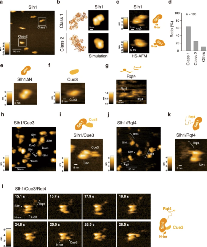
a HS-AFM image of Slh1. Two major particles were indicated as Class1 and Class2. b The pseudo-AFM images of Slh1 belonging to Class1 and Class2 particles, which were simulated using predicted Slh1 structure lacking N-terminal region by Alphafold2. c The HS-AFM images and schematized molecular features of Slh1. d Classification of Slh1 particles. All particles used for the classification were presented in the supplementary Fig. 4. e HS-AFM images of Slh1 lacking N-terminal region (Slh1∆N). f HS-AFM image of Cue3. g HS-AFM image of Rqt4. h, i HS-AFM images of Slh1/Cue3 complex. j, k HS-AFM images of Slh1/Rqt4 complex. l The time-lapse HS-AFM images of the RQT complex. All experiments were performed at least twice with highly reproducible results.

This article beautifully merges state-of-the-art structural biology with nanotechnology tools to reveal how molecular collisions are detected and resolved. The incorporation of ultra-fast AFM probe technology makes it possible to observe RQT in action, delivering new insight into cellular quality control.

Real-time multistep asymmetrical disassembly of nucleosomes and chromatosomes visualized by high-speed atomic force microscopy

During replication, expression, and repair of the eukaryotic genome, cellular machinery must access the DNA wrapped around histone proteins forming nucleosomes. These octameric protein·DNA complexes are modular, dynamic, and flexible and unwrap or disassemble either spontaneously or by the action of molecular motors. Thus, the mechanism of formation and regulation of subnucleosomal intermediates has gained attention genome-wide because it controls DNA accessibility.*

In the article  “Real-Time Multistep Asymmetrical Disassembly of Nucleosomes and Chromatosomes Visualized by High-Speed Atomic Force Microscopy” Bibiana Onoa, César Díaz-Celis, Cristhian Cañari-Chumpitaz, Antony Lee and Carlos Bustamante describe how they imaged nucleosomes and their more compacted structure with the linker histone H1 (chromatosomes) using high-speed atomic force microscopy to visualize simultaneously the changes in the DNA and the histone core during their disassembly when deposited on mica.*

Furthermore, Bibiana Onoa et al. trained a neural network and developed an automatic algorithm to track molecular structural changes in real time. *

The authors’ results show that nucleosome disassembly is a sequential process involving asymmetrical stepwise dimer ejection events. The presence of H1 restricts DNA unwrapping, significantly increases the nucleosomal lifetime, and affects the pathway in which heterodimer asymmetrical dissociation occurs. *

Bibiana Onoa et al.  observe that tetrasomes are resilient to disassembly and that the tetramer core (H3·H4)2 can diffuse along the nucleosome positioning sequence. Tetrasome mobility might be critical to the proper assembly of nucleosomes and can be relevant during nucleosomal transcription, as tetrasomes survive RNA polymerase passage. These findings are relevant to understanding nucleosome intrinsic dynamics and their modification by DNA-processing enzymes. *

To characterize the nucleosomes dynamics in 2D, individual molecules were observed in buffer using an Ando-type high speed atomic force microscope together with NanoWorld Ultra-Short Cantilevers for HS-AFM of the USC-F1.2-K0.15 AFM probe type ( typical spring constant 0.15 N/m, typical resonance frequency in air 1200 kHz, resonance frequency 500–600 kHz in liquid). *

The AFM data presented in the article allow the authors to directly visualize the dynamics of DNA and histones during nucleosome and chromatosome disassembly, providing a simultaneous observation of DNA unwrapping and histone dissociation. *

The experimental and analytical strategy presented shows that real-time HS-AFM is a robust and powerful tool for studying single nucleosomes and chromatin dynamics. *

graphical abstract from Bibiana Onoa et al 2024 "Real-Time Multistep Asymmetrical Disassembly of Nucleosomes and Chromatosomes Visualized by High-Speed Atomic Force Microscopy" - NanoWorld Ultra-Short Cantilevers of the USC-F1.2-k0.15 AFM probe type were used for the high-speed atomic force microscopy
graphical abstract from Bibiana Onoa et al 2024 “Real-Time Multistep Asymmetrical Disassembly of Nucleosomes and Chromatosomes Visualized by High-Speed Atomic Force Microscopy”

*Bibiana Onoa, César Díaz-Celis, Cristhian Cañari-Chumpitaz, Antony Lee and Carlos Bustamante
Real-Time Multistep Asymmetrical Disassembly of Nucleosomes and Chromatosomes Visualized by High-Speed Atomic Force Microscopy
ACS Central Science 2024, 10, 1, 122–137
DOI: https://doi.org/10.1021/acscentsci.3c00735

Open Access The article “Real-Time Multistep Asymmetrical Disassembly of Nucleosomes and Chromatosomes Visualized by High-Speed Atomic Force Microscopy” by Bibiana Onoa, César Díaz-Celis, Cristhian Cañari-Chumpitaz, Antony Lee and Carlos Bustamante is licensed under a Creative Commons Attribution 4.0 International License, which permits use, sharing, adaptation, distribution and reproduction in any medium or format, as long as you give appropriate credit to the original author(s) and the source, provide a link to the Creative Commons license, and indicate if changes were made. The images or other third party material in this article are included in the article’s Creative Commons license, unless indicated otherwise in a credit line to the material. If material is not included in the article’s Creative Commons license and your intended use is not permitted by statutory regulation or exceeds the permitted use, you will need to obtain permission directly from the copyright holder. To view a copy of this license, visit http://creativecommons.org/licenses/by/4.0/.

Intrinsically disordered regions in TRPV2 mediate protein-protein interactions

Transient receptor potential (TRP) ion channels are gated by diverse intra- and extracellular stimuli leading to cation inflow (Na+, Ca2+) regulating many cellular processes and initiating organismic somatosensation. *

Structures of most TRP channels have been solved. However, structural and sequence analysis showed that ~30% of the TRP channel sequences, mainly the N- and C-termini, are intrinsically disordered regions (IDRs). Unfortunately, very little is known about IDR ‘structure’, dynamics and function, though it has been shown that they are essential for native channel function. *

In the article “Intrinsically disordered regions in TRPV2 mediate protein-protein interactions”, Raghavendar R. Sanganna Gari, Grigory Tagiltsev, Ruth A. Pumroy, Yining Jiang, Martin Blackledge, Vera Y. Moiseenkova-Bell and Simon Scheuring imaged TRPV2 channels in membranes using high-speed atomic force microscopy (HS-AFM). *

The dynamic single molecule imaging capability of HS-AFM allowed the authors to visualize IDRs and revealed that N-terminal IDRs were involved in intermolecular interactions. Their work provides evidence about the ‘structure’ of the TRPV2 IDRs, and that the IDRs may mediate protein-protein interactions. *

In total, 1.5 µl of the TRPV2 reconstituted vesicles were deposited on a 1.5-mm2 freshly cleaved mica surface, which was glued with epoxy to the quartz sample stage. After 20–30 min incubation, the sample was gently rinsed with imaging buffer (20 mM Hepes, pH 8.0, 150 mM NaCl) and mounted in the HS-AFM fluid cell. All images in this study were taken using a HS-AFM operated in amplitude modulation mode using optimized scan and feedback parameters and lab-built amplitude detectors and free amplitude stabilizers. *

Short (8 µm) cantilevers (NanoWorld Ultra-Short Cantilevers for High-Speed AFM USC-F1.2-k0.15) with nominal spring constant of 0.15 N/m, resonance frequency of 0.6 MHz, and a quality factor of ∼1.5 in liquid were used. AFM probes were sharpened using oxygen plasma etching to obtain better resolution. *

Fig. 1 from “Intrinsically disordered regions in TRPV2 mediate protein-protein interactions” by Raghavendar R. Sanganna Gari et al. :TRPV2 reconstitution for HS-AFM analysis. b Overview HS-AFM images (Supplementary Movie 1) of TRPV2 (windmill-shaped molecules) in soy polar lipid membranes on mica (dark background areas). False color scale: 0–9 nm. The white oversaturated areas have a height of ~26 nm and represent likely non-ruptured small vesicles. NanoWorld-USC-F1.2-k0.15 AFM probes were used for the HS-AFM
Fig. 1 from “Intrinsically disordered regions in TRPV2 mediate protein-protein interactions” by Raghavendar R. Sanganna Gari et al. :
TRPV2 reconstitution for HS-AFM analysis.
b Overview HS-AFM images (Supplementary Movie 1) of TRPV2 (windmill-shaped molecules) in soy polar lipid membranes on mica (dark background areas). False color scale: 0–9 nm. The white oversaturated areas have a height of ~26 nm and represent likely non-ruptured small vesicles.
Fig. 1 from “Intrinsically disordered regions in TRPV2 mediate protein-protein interactions” by Raghavendar R. Sanganna Gari et al. : TRPV2 reconstitution for HS-AFM analysis. a Negative-stain EM of TRPV2 reconstituted into soy polar lipids at a lipid-to-protein ratio of 0.7. Protruding features (arrow) at the vesicle periphery and the strong contrast of the proteins in the vesicle in the negative-stain EM are indicative of inside-out reconstitution of the TRPV2 channels with the large cytoplasmic domains exposed to the outside of the vesicle. b Overview HS-AFM images (Supplementary Movie 1) of TRPV2 (windmill-shaped molecules) in soy polar lipid membranes on mica (dark background areas). False color scale: 0–9 nm. The white oversaturated areas have a height of ~26 nm and represent likely non-ruptured small vesicles. c Height distribution of TRPV2 above mica from (b). TRPV2 has a full height of 9.5 ± 0.1 nm above mica, in good agreement with the TRPV2 cryo-EM structure. Inset: Cryo-EM structure PDB 6U84 shown with the intracellular side up (as imaged by HS-AFM), membrane indicated in light gray. Short (8 µm) cantilevers (NanoWorld Ultra-Short Cantilevers for High-Speed AFM USC-F1.2-k0.15,) with nominal spring constant of 0.15 N/m, resonance frequency of 0.6 MHz, and a quality factor of ∼1.5 in liquid were used. AFM probes were sharpened using oxygen plasma etching to obtain better resolution. *
Fig. 1 from “Intrinsically disordered regions in TRPV2 mediate protein-protein interactions” by Raghavendar R. Sanganna Gari et al. :
TRPV2 reconstitution for HS-AFM analysis.
a Negative-stain EM of TRPV2 reconstituted into soy polar lipids at a lipid-to-protein ratio of 0.7. Protruding features (arrow) at the vesicle periphery and the strong contrast of the proteins in the vesicle in the negative-stain EM are indicative of inside-out reconstitution of the TRPV2 channels with the large cytoplasmic domains exposed to the outside of the vesicle. b Overview HS-AFM images (Supplementary Movie 1) of TRPV2 (windmill-shaped molecules) in soy polar lipid membranes on mica (dark background areas). False color scale: 0–9 nm. The white oversaturated areas have a height of ~26 nm and represent likely non-ruptured small vesicles. c Height distribution of TRPV2 above mica from (b). TRPV2 has a full height of 9.5 ± 0.1 nm above mica, in good agreement with the TRPV2 cryo-EM structure. Inset: Cryo-EM structure PDB 6U84 shown with the intracellular side up (as imaged by HS-AFM), membrane indicated in light gray.


*Raghavendar R. Sanganna Gari, Grigory Tagiltsev, Ruth A. Pumroy, Yining Jiang, Martin Blackledge, Vera Y. Moiseenkova-Bell and Simon Scheuring
Intrinsically disordered regions in TRPV2 mediate protein-protein interactions
Communications Biology volume 6, Article number: 966 (2023)
DOI: https://doi.org/10.1038/s42003-023-05343-7

Please follow this external link to read the full article: https://rdcu.be/dnNba

The article “Phosphorylation of phase-separated p62 bodies by ULK1 activates a redox-independent stress response” by Raghavendar R. Sanganna Gari, Grigory Tagiltsev, Ruth A. Pumroy, Yining Jiang, Martin Blackledge, Vera Y. Moiseenkova-Bell and Simon Scheuring is licensed under a Creative Commons Attribution 4.0 International License, which permits use, sharing, adaptation, distribution and reproduction in any medium or format, as long as you give appropriate credit to the original author(s) and the source, provide a link to the Creative Commons license, and indicate if changes were made. The images or other third-party material in this article are included in the article’s Creative Commons license, unless indicated otherwise in a credit line to the material. If material is not included in the article’s Creative Commons license and your intended use is not permitted by statutory regulation or exceeds the permitted use, you will need to obtain permission directly from the copyright holder. To view a copy of this license, visit https://creativecommons.org/licenses/by/4.0/.