
Controllable Gliders in a Nanomagnetic Metamaterial: MFM Reveals Magnetic “Snakes”

Artificial Spin Ice (ASI) has emerged as a fascinating platform for studying collective behaviour in interacting nanomagnets, with potential applications ranging from magnetic devices to neuromorphic computing. In this article, Arthur Penty, Johannes H. Jensen, Ida Breivik, Anders Strømberg, Erik Folven, and Gunnar Tufte demonstrate how a mobile magnetic structure — a “snake” glider — can be created and precisely controlled within a nanomagnetic metamaterial.

Inspired by the concept of gliders in Cellular Automata, the researchers use an evolutionary algorithm to discover a simple moving structure within a pinwheel Artificial Spin Ice system. The resulting snake can move through the nanomagnetic array under a global magnetic-field protocol, allowing controlled manipulation of magnetic textures on a scale of approximately 100 nm.

Artificial Spin Ice as a Platform for Information Processing

Artificial Spin Ice consists of interacting nanomagnets arranged in a two-dimensional lattice. These nanoscale magnetic elements can exhibit collective states and emergent behaviour, making ASI attractive for exploring unconventional approaches to information processing.

A key challenge is integrating information transformation, transmission, and storage into the same physical system. The snake glider introduced in this article addresses this challenge by providing a controllable magnetic structure that can move through the ASI lattice while retaining its functionality.

The researchers demonstrate that the snake can move either left or right depending on its orientation. Its motion is controlled through a sequence of in-plane magnetic fields, while simulations and experiments are used to investigate the mechanism responsible for the glider’s movement and its robustness against disorder.

NanoWorld MFMR for Magnetic Force Microscopy

Experimental investigation of the nanomagnetic structures was performed using NanoWorld POINTPROBE MFMR, a magnetic force microscopy AFM probe designed for magnetic imaging.

The NanoWorld MFMR was used for both writing the initial magnetic state and imaging the resulting magnetic structures. To initialise the snake, the MFM probe was used to write an approximately 1 µm magnetic line in the pinwheel Artificial Spin Ice while a 10 mT bias field was applied. The writing process was performed with the MFM probe in contact with the sample at a scan speed of 55 µm/s.
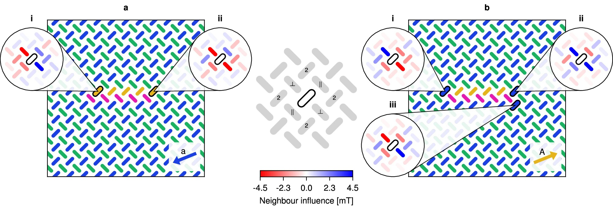
Fig. 5: Analysis of neighbour influence in the snake.
Fig. 5: Analysis of neighbour influence in the snake. Neighbour influence during (a) shrinking and (b) growth. The five insets illustrate how the highlighted magnet is influenced by its neighbours through their dipolar fields. The influence of a neighbour acting on a magnet is given by the resulting change in the magnet’s proximity to the switching astroid when considering the neighbour’s dipolar field. Positive influence values (blue) indicate an increased distance (stabilising) and negative values (red) indicate a decreased distance (destabilising). The centre legend shows the parallel (∥) and perpendicular (⊥) nearest neighbours, as well as the second nearest neighbours (2).

Following magnetic-field-driven evolution of the structure, the researchers used MFM to image the magnetic state after each applied field. Imaging was performed at remanence using a lift height between 55 and 60 nm and scan speeds between 50 and 55 µm/s. All experiments were conducted at room temperature.

These measurements allowed the researchers to directly observe how the magnetic texture evolved as the snake moved through the nanomagnetic array.

Imaging a Controllable Magnetic Texture

The experimental MFM results provide a direct view of the magnetic states underlying the glider behaviour. By repeatedly applying the field protocol and imaging the resulting configurations, the researchers could track the movement of individual snakes and investigate how their structure responds to the magnetic environment.

The experiments also demonstrated the robustness of the concept. Multiple snakes could be initialised within the same Artificial Spin Ice array while maintaining sufficient separation to avoid unwanted interactions. The researchers further investigated how fabrication disorder affects the movement of the gliders, finding evidence of self-correction and graceful degradation before motion eventually breaks down.

The combination of experimental MFM imaging and micromagnetic simulations provides a detailed picture of the mechanism behind the snake’s movement. This makes the approach particularly interesting for studying how local magnetic interactions can be harnessed to produce controlled, collective behaviour.

From Magnetic Gliders to Neuromorphic Computing

The snake glider provides more than a visually compelling magnetic structure. Because its position and movement can be controlled, it can act as a physical carrier of information within the Artificial Spin Ice substrate.

The researchers demonstrate how the glider can contribute to information transmission, storage, and transformation, potentially allowing these functions to be integrated into a single magnetic material. Such concepts could contribute to the development of ultra-low-power computing architectures based on nanoscale magnetic states rather than conventional electronic switching.

For this type of research, high-quality MFM probes are

essential for resolving and manipulating nanoscale magnetic structures. The NanoWorld POINTPROBE MFMR provides a dedicated solution for magnetic force microscopy, enabling both magnetic writing and high-resolution imaging of nanomagnetic systems.

The work by Penty and co-authors demonstrates how MFM can do more than simply characterise magnetic materials: it can become an experimental tool for exploring programmable magnetic textures, emergent behaviour, and new approaches to information processing.

Full citation:
Penty, A.; Jensen, J. H.; Breivik, I.; Strømberg, A.; Folven, E.; Tufte, G.
Controllable gliders in a nanomagnetic metamaterial.
Nature Communications 2025, 16, 7500.
DOI: 10.1038/s41467-025-62515-1

License: CC BY 4.0

Bi2Se3 interlayer treatments affecting the Y3Fe5O12 (YIG) platinum spin Seebeck effect

Spin Seebeck effects (SSE) arise from spin current (magnon) generation from within ferri-, ferro-, or anti-ferromagnetic materials driven by an applied temperature gradient. *

Longitudinal spin Seebeck effect (LSSE) investigations, where the spin current and temperature gradient evolve along a common z axis, while the magnetic field is applied in the y axis and the voltage contacts are spaced along the x axis, have become the most popular spin Seebeck device architecture. *

In article “Bi2Se3 interlayer treatments affecting the Y3Fe5O12 (YIG) platinum spin Seebeck effect”, Yaoyang Hu, Michael P. Weir, H. Jessica Pereira, Oliver J. Amin, Jem Pitcairn, Matthew J. Cliffe, Andrew W. Rushforth, Gunta Kunakova, Kiryl Niherysh, Vladimir Korolkov, James Kertfoot, Oleg Makarovsky and Simon Woodward present a method to enhance the longitudinal spin Seebeck effect at platinum/yttrium iron garnet (Pt/YIG) interfaces. *

The introduction of a partial interlayer of bismuth selenide (Bi2Se3, 2.5% surface coverage) interfaces significantly increases (by ∼380%–690%) the spin Seebeck coefficient over equivalent Pt/YIG control devices. *

Optimal devices are prepared by transferring Bi2Se3 nanoribbons, prepared under anaerobic conditions, onto the YIG (111) chips followed by rapid over-coating with Pt. The deposited Pt/Bi2Se3 nanoribbon/YIG assembly is characterized by scanning electron microscope. The expected elemental compositions of Bi2Se3 and YIG are confirmed by energy dispersive x-ray analysis. *

A spin Seebeck coefficient of 0.34–0.62 μV/K for Pt/Bi2Se3/YIG is attained for the authors’ devices, compared to just 0.09 μV/K for Pt/YIG controls at a 12 K thermal gradient and a magnetic field swept from −50 to +50 mT. *

Superconducting quantum interference device magnetometer studies indicate that the magnetic moment of Pt/Bi2Se3/YIG treated chips is increased by ∼4% vs control Pt/YIG chips (i.e., a significant increase vs the ±0.06% chip mass reproducibility). *

Increased surface magnetization is also detected in magnetic force microscope studies of Pt/Bi2Se3/YIG, suggesting that the enhancement of spin injection is associated with the presence of Bi2Se3 nanoribbons. *

To understand the surface magnetization effects in sample BSYIG1-a further, magnetic force microscope (MFM) measurements were undertaken using a commercial atomic force microscope and magnetic NanoWorld Pointprobe® MFMR AFM probes. *

MFM differs from traditional atomic force microscopy in that the AFM probe, in addition to providing a surface height profile, is also able to detect the magnetic field gradient above the sample. *

MFM surface profiling of BSYIG1-a revealed that a typical ribbon is comprised of multilayers of Bi2Se3, providing thicker sections ca. 250 nm thick [e.g., the profile along vector 1 in Figs. 3(a) and 3(b) cited below] and additional thinner sections ca. 100 nm thick [e.g., the profile along vector 2 in Figs. 3(a) and 3(b)]. Re-running ribbon profiles 1 and 2 with the magnetic probe at a height of 100 nm above the topological surface provided data on the magnetic field gradient variation along the same line profiles. The MFM amplitude [Figs. 3(c) and 3(d) cited below] increases over the Bi2Se3 flake, and furthermore, the magnetic enhancement correlates with the thickness of the Bi2Se3, being larger for the thicker part of the sample. *

This amplitude enhancement suggests that the observed effect is magnetic rather than due to long-range electrostatics, supporting the inference that the surface magnetization is improved by the presence of Bi2Se3 flakes at the interlayer of a Pt/YIG device. However, it was not possible to extract quantitative information about surface magnetization from this study, but Yaoyang Hu et al. are hopeful that future experimental and theoretical work can provide further explanation. *

Figure 3 from Yaoyang Hu et al. “Bi2Se3 interlayer treatments affecting the Y3Fe5O12 (YIG) platinum spin Seebeck effect”:Scanning probe microscopy images of BSYIG1-a: (a) Atomic force microscopy image of a representative Bi2Se3 nanoribbon on a YIG/GGG substrate. (b) Bi2Se3 ribbon profile scans along vectors 1 (pink) and 2 (blue) showing the two differential height responses. (c) Magnetic force microscopy image of the same Bi2Se3 nanoribbon. The measurement was performed at 100 nm above the topological heights determined in the AFM study. (d) MFM profile scans along vectors 1 (pink) and 2 (blue) showing the magnetic response. Magnetic force microscope (MFM) measurements were undertaken using a commercial atomic force microscope and magnetic NanoWorld MFMR AFM probes. *
Figure 3 from Yaoyang Hu et al. “Bi2Se3 interlayer treatments affecting the Y3Fe5O12 (YIG) platinum spin Seebeck effect”:
Scanning probe microscopy images of BSYIG1-a: (a) Atomic force microscopy image of a representative Bi2Se3 nanoribbon on a YIG/GGG substrate. (b) Bi2Se3 ribbon profile scans along vectors 1 (pink) and 2 (blue) showing the two differential height responses. (c) Magnetic force microscopy image of the same Bi2Se3 nanoribbon. The measurement was performed at 100 nm above the topological heights determined in the AFM study. (d) MFM profile scans along vectors 1 (pink) and 2 (blue) showing the magnetic response.

*Yaoyang Hu, Michael P. Weir, H. Jessica Pereira, Oliver J. Amin, Jem Pitcairn, Matthew J. Cliffe, Andrew W. Rushforth, Gunta Kunakova, Kiryl Niherysh, Vladimir Korolkov, James Kertfoot, Oleg Makarovsky and Simon Woodward
Bi2Se3 interlayer treatments affecting the Y3Fe5O12 (YIG) platinum spin Seebeck effect
Applied Physics Letters 123, 223902 (2023)
DOI: https://doi.org/10.1063/5.0157778

The article “Bi2Se3 interlayer treatments affecting the Y3Fe5O12 (YIG) platinum spin Seebeck effect” by Yaoyang Hu, Michael P. Weir, H. Jessica Pereira, Oliver J. Amin, Jem Pitcairn, Matthew J. Cliffe, Andrew W. Rushforth, Gunta Kunakova, Kiryl Niherysh, Vladimir Korolkov, James Kertfoot, Oleg Makarovsky and Simon Woodward is licensed under a Creative Commons Attribution 4.0 International License, which permits use, sharing, adaptation, distribution and reproduction in any medium or format, as long as you give appropriate credit to the original author(s) and the source, provide a link to the Creative Commons license, and indicate if changes were made. The images or other third-party material in this article are included in the article’s Creative Commons license, unless indicated otherwise in a credit line to the material. If material is not included in the article’s Creative Commons license and your intended use is not permitted by statutory regulation or exceeds the permitted use, you will need to obtain permission directly from the copyright holder. To view a copy of this license, visit https://creativecommons.org/licenses/by/4.0/.

Nanoscale Noncollinear Spin Textures in Thin Films of a D2d Heusler Compound

Magnetic nano-objects, namely antiskyrmions and Bloch skyrmions, have been found to coexist in single-crystalline lamellae formed from bulk crystals of inverse tetragonal Heusler compounds with D2d symmetry. *

Skyrmions can be observed in real-space by various direct imaging techniques. *

In the article “Nanoscale Noncollinear Spin Textures in Thin Films of a D2d Heusler Compound”  Ankit K. Sharma, Jagannath Jena, Kumari Gaurav Rana, Anastasios Markou, Holger L. Meyerheim, Katayoon Mohseni, Abhay K. Srivastava, Ilya Kostanoskiy, Claudia Felser and Stuart S. P. Parkin  describe the use of magnetic force microscopy (MFM) imaging to investigate magnetic textures in epitaxial thin films of [001]-oriented Mn2RhSn formed by magnetron sputtering.*

They find evidence for magnetic nano-objects which exhibit a wide range of sizes with stability with respect to magnetic field and temperature that is similar to single-crystalline lamellae. *

However, the nano-objects do not form well-defined arrays, nor is any evidence found for helical spin textures. This is speculated to likely be a consequence of the poorer homogeneity of chemical ordering in the thin films. *

Evidence is found for elliptically distorted nano-objects along perpendicular crystallographic directions within the epitaxial films, which is consistent with elliptical Bloch skyrmions observed in single-crystalline lamellae. Thus, these measurements provide strong evidence for the formation of noncollinear spin textures in thin films of Mn2RhSn. *

Using these films, it is shown that individual nano-objects can be deleted using a local magnetic field from a magnetic AFM tip and collections of nano-objects can be similarly written. *

For writing and deleting the nano-objects, magnetic AFM probes from NanoWorld of the Pointprobe® MFMR type were used. *

These observations described in the article suggest a path toward the use of these nano-objects in thin films with D2d symmetry as magnetic memory elements paving the way to the realization of skyrmionic devices based on Heusler thin films. *

Figure 5 from Ankit K. Sharma et al. Nanoscale Noncollinear Spin Textures in Thin Films of a D2d Heusler Compound : Controlled creation and annihilation of nano-objects in a 35 nm-thick Mn2RhSn thin film. a) Schematic illustration of magnetization orientations of MFM tip and sample for writing. The distance between tip and the sample is the scan height z. b–e) MFM images in zero field and z = 80, 40, 30, and 20 nm, respectively at 200 K. f) Contact-mode image in zero field and 200 K. The blue and red colors represent up and down magnetization, respectively. Images in (b)–(f) are at the same scale: a scale bar is given in (f). g–i) MFM images taken at z = 70, 50, and 30 nm at 100 K under Hz = 180 mT. Images in (g)–(i) are at the same scale: a scale bar is given in (i). For writing and deleting the nano-objects, magnetic AFM probes from NanoWorld of the Pointprobe® MFMR type were used.
Figure 5 from Ankit K. Sharma et al. Nanoscale Noncollinear Spin Textures in Thin Films of a D2d Heusler Compound :
Controlled creation and annihilation of nano-objects in a 35 nm-thick Mn2RhSn thin film. a) Schematic illustration of magnetization orientations of MFM tip and sample for writing. The distance between tip and the sample is the scan height z. b–e) MFM images in zero field and z = 80, 40, 30, and 20 nm, respectively at 200 K. f) Contact-mode image in zero field and 200 K. The blue and red colors represent up and down magnetization, respectively. Images in (b)–(f) are at the same scale: a scale bar is given in (f). g–i) MFM images taken at z = 70, 50, and 30 nm at 100 K under Hz = 180 mT. Images in (g)–(i) are at the same scale: a scale bar is given in (i).

*Ankit K. Sharma, Jagannath Jena, Kumari Gaurav Rana, Anastasios Markou, Holger L. Meyerheim, Katayoon Mohseni, Abhay K. Srivastava, Ilya Kostanoskiy, Claudia Felser, Stuart S. P. Parkin
Nanoscale Noncollinear Spin Textures in Thin Films of a D2d Heusler Compound
Advanced Materials, Volume 33, Issue 32, August 12, 2021, 2101323
DOI: https://doi.org/10.1002/adma.202101323

Open Access The article “Nanoscale Noncollinear Spin Textures in Thin Films of a D2d Heusler Compound” Ankit K. Sharma, Jagannath Jena, Kumari Gaurav Rana, Anastasios Markou, Holger L. Meyerheim, Katayoon Mohseni, Abhay K. Srivastava, Ilya Kostanoskiy, Claudia Felser and Stuart S. P. Parkin is licensed under a Creative Commons Attribution 4.0 International License, which permits use, sharing, adaptation, distribution and reproduction in any medium or format, as long as you give appropriate credit to the original author(s) and the source, provide a link to the Creative Commons license, and indicate if changes were made. The images or other third party material in this article are included in the article’s Creative Commons license, unless indicated otherwise in a credit line to the material. If material is not included in the article’s Creative Commons license and your intended use is not permitted by statutory regulation or exceeds the permitted use, you will need to obtain permission directly from the copyright holder. To view a copy of this license, visit http://creativecommons.org/licenses/by/4.0/.