
Controllable Gliders in a Nanomagnetic Metamaterial: MFM Reveals Magnetic “Snakes”

Artificial Spin Ice (ASI) has emerged as a fascinating platform for studying collective behaviour in interacting nanomagnets, with potential applications ranging from magnetic devices to neuromorphic computing. In this article, Arthur Penty, Johannes H. Jensen, Ida Breivik, Anders Strømberg, Erik Folven, and Gunnar Tufte demonstrate how a mobile magnetic structure — a “snake” glider — can be created and precisely controlled within a nanomagnetic metamaterial.

Inspired by the concept of gliders in Cellular Automata, the researchers use an evolutionary algorithm to discover a simple moving structure within a pinwheel Artificial Spin Ice system. The resulting snake can move through the nanomagnetic array under a global magnetic-field protocol, allowing controlled manipulation of magnetic textures on a scale of approximately 100 nm.

Artificial Spin Ice as a Platform for Information Processing

Artificial Spin Ice consists of interacting nanomagnets arranged in a two-dimensional lattice. These nanoscale magnetic elements can exhibit collective states and emergent behaviour, making ASI attractive for exploring unconventional approaches to information processing.

A key challenge is integrating information transformation, transmission, and storage into the same physical system. The snake glider introduced in this article addresses this challenge by providing a controllable magnetic structure that can move through the ASI lattice while retaining its functionality.

The researchers demonstrate that the snake can move either left or right depending on its orientation. Its motion is controlled through a sequence of in-plane magnetic fields, while simulations and experiments are used to investigate the mechanism responsible for the glider’s movement and its robustness against disorder.

NanoWorld MFMR for Magnetic Force Microscopy

Experimental investigation of the nanomagnetic structures was performed using NanoWorld POINTPROBE MFMR, a magnetic force microscopy AFM probe designed for magnetic imaging.

The NanoWorld MFMR was used for both writing the initial magnetic state and imaging the resulting magnetic structures. To initialise the snake, the MFM probe was used to write an approximately 1 µm magnetic line in the pinwheel Artificial Spin Ice while a 10 mT bias field was applied. The writing process was performed with the MFM probe in contact with the sample at a scan speed of 55 µm/s.
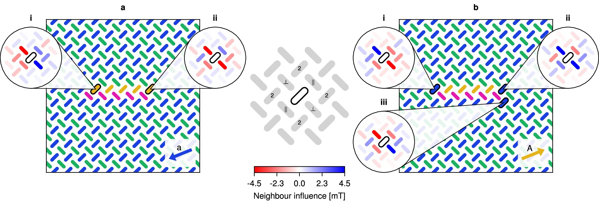
Fig. 5: Analysis of neighbour influence in the snake.
Fig. 5: Analysis of neighbour influence in the snake. Neighbour influence during (a) shrinking and (b) growth. The five insets illustrate how the highlighted magnet is influenced by its neighbours through their dipolar fields. The influence of a neighbour acting on a magnet is given by the resulting change in the magnet’s proximity to the switching astroid when considering the neighbour’s dipolar field. Positive influence values (blue) indicate an increased distance (stabilising) and negative values (red) indicate a decreased distance (destabilising). The centre legend shows the parallel (∥) and perpendicular (⊥) nearest neighbours, as well as the second nearest neighbours (2).

Following magnetic-field-driven evolution of the structure, the researchers used MFM to image the magnetic state after each applied field. Imaging was performed at remanence using a lift height between 55 and 60 nm and scan speeds between 50 and 55 µm/s. All experiments were conducted at room temperature.

These measurements allowed the researchers to directly observe how the magnetic texture evolved as the snake moved through the nanomagnetic array.

Imaging a Controllable Magnetic Texture

The experimental MFM results provide a direct view of the magnetic states underlying the glider behaviour. By repeatedly applying the field protocol and imaging the resulting configurations, the researchers could track the movement of individual snakes and investigate how their structure responds to the magnetic environment.

The experiments also demonstrated the robustness of the concept. Multiple snakes could be initialised within the same Artificial Spin Ice array while maintaining sufficient separation to avoid unwanted interactions. The researchers further investigated how fabrication disorder affects the movement of the gliders, finding evidence of self-correction and graceful degradation before motion eventually breaks down.

The combination of experimental MFM imaging and micromagnetic simulations provides a detailed picture of the mechanism behind the snake’s movement. This makes the approach particularly interesting for studying how local magnetic interactions can be harnessed to produce controlled, collective behaviour.

From Magnetic Gliders to Neuromorphic Computing

The snake glider provides more than a visually compelling magnetic structure. Because its position and movement can be controlled, it can act as a physical carrier of information within the Artificial Spin Ice substrate.

The researchers demonstrate how the glider can contribute to information transmission, storage, and transformation, potentially allowing these functions to be integrated into a single magnetic material. Such concepts could contribute to the development of ultra-low-power computing architectures based on nanoscale magnetic states rather than conventional electronic switching.

For this type of research, high-quality MFM probes are

essential for resolving and manipulating nanoscale magnetic structures. The NanoWorld POINTPROBE MFMR provides a dedicated solution for magnetic force microscopy, enabling both magnetic writing and high-resolution imaging of nanomagnetic systems.

The work by Penty and co-authors demonstrates how MFM can do more than simply characterise magnetic materials: it can become an experimental tool for exploring programmable magnetic textures, emergent behaviour, and new approaches to information processing.

Full citation:
Penty, A.; Jensen, J. H.; Breivik, I.; Strømberg, A.; Folven, E.; Tufte, G.
Controllable gliders in a nanomagnetic metamaterial.
Nature Communications 2025, 16, 7500.
DOI: 10.1038/s41467-025-62515-1

License: CC BY 4.0

NanoWorld at MRS Fall Meeting & Exhibit 2023 this week

NanoWorld CEO Manfred Detterbeck is in Boston for the MRS Fall 2023 Meeting & Exhibit this week. Will you be there too? You’ll meet him at some of the sessions or at NanoAndMore USA booth no. 904.

NanoWorld AFM probes CEO Manfred Detterbeck pointing at the MRS Fall 2023 Meeting & Exhibit sign.
NanoWorld AG CEO Manfred Detterbeck at MRS Fall 2023

Boron nitride nanoresonators for phonon-enhanced molecular vibrational spectroscopy at the strong coupling limit

In the article “Boron nitride nanoresonators for phonon-enhanced molecular vibrational spectroscopy at the strong coupling limit” the authors use, for the first time, phonon-polariton-resonant h-BN ribbons for SEIRA spectroscopy of small amounts of organic molecules in Fourier transform infrared spectroscopy. They demonstrate a new way to strongly couple infrared light and molecular vibrations, by utilizing phonon polariton nanoresonators made of hexagonal boron nitride, a Van der Waals material.

For the nanoscale Fourier transform infrared (nano-FTIR) spectroscopy mentioned in this article an oscillating Pt/Ir coated NanoWorld Arrow-NCPt AFM probe was illuminated by p-polarized mid-IR broadband radiation.

Figure 2 from "Boron nitride nanoresonators for phonon-enhanced molecular vibrational spectroscopy at the strong coupling limit": Far- and near-field spectroscopic characterization of h-BN ribbon arrays. (a) Sketch of the transmission spectroscopy experiment. Incoming light at normal incidence is polarized perpendicular to the ribbons to excite the HPhP resonance. (b) Transmission spectrum normalized to the bare substrate spectrum, T/T0, for a 20 × 20 μm2 h-BN ribbon array. Ribbon width w=158 nm, ribbon period D=400 nm and ribbon height h=40 nm. (c) Sketch of the nano-FTIR spectroscopy experiment. The near-field probing tip is scanned across (y-direction) the h-BN ribbon in 20-nm steps, as indicated by the dashed blue line. Near-field spectra are recorded as a function of the tip position (the detector signal is demodulated at the third harmonic of the tip tapping frequency, yielding s3(y, ω), as explained in the Materials and methods section). (d) Lower panel: Spectral line scan s3(y, ω), where each horizontal line corresponds to a spectrum recorded at a fixed y-position (vertical axis). Upper panel: Illustration of the real part of the z-component of the electric field (Re[Ez]) profile across the ribbon at the resonance frequency observed in the nano-FTIR spectra (lower panel). The AFM tip used was a NanoWorld Arrow-NCPT
Figure 2 from “Boron nitride nanoresonators for phonon-enhanced molecular vibrational spectroscopy at the strong coupling limit”: Far- and near-field spectroscopic characterization of h-BN ribbon arrays. (a) Sketch of the transmission spectroscopy experiment. Incoming light at normal incidence is polarized perpendicular to the ribbons to excite the HPhP resonance. (b) Transmission spectrum normalized to the bare substrate spectrum, T/T0, for a 20 × 20 μm2 h-BN ribbon array. Ribbon width w=158 nm, ribbon period D=400 nm and ribbon height h=40 nm. (c) Sketch of the nano-FTIR spectroscopy experiment. The near-field probing tip is scanned across (y-direction) the h-BN ribbon in 20-nm steps, as indicated by the dashed blue line. Near-field spectra are recorded as a function of the tip position (the detector signal is demodulated at the third harmonic of the tip tapping frequency, yielding s3(y, ω), as explained in the Materials and methods section). (d) Lower panel: Spectral line scan s3(y, ω), where each horizontal line corresponds to a spectrum recorded at a fixed y-position (vertical axis). Upper panel: Illustration of the real part of the z-component of the electric field (Re[Ez]) profile across the ribbon at the resonance frequency observed in the nano-FTIR spectra (lower panel).


Marta Autore, Peining Li, Irene Dolado, Francisco J Alfaro-Mozaz, Ruben Esteban, Ainhoa Atxabal, Fèlix Casanova, Luis E Hueso, Pablo Alonso-González, Javier Aizpurua, Alexey Y Nikitin, Saül Vélez & Rainer Hillenbrand
Boron nitride nanoresonators for phonon-enhanced molecular vibrational spectroscopy at the strong coupling limit
Light: Science & Applications volume 7, page 17172 (2018)
DOI: https://doi.org/10.1038/lsa.2017.172

For the full article please follow this external link: https://rdcu.be/7B0F

The article: Boron nitride nanoresonators for phonon-enhanced molecular vibrational spectroscopy at the strong coupling limit by Marta Autore et. al, is licensed under a Creative Commons Attribution 4.0 International License. The images or other third party material in this article are included in the article’s Creative Commons license, unless indicated otherwise in the credit line; if the material is not included under the Creative Commons license, users will need to obtain permission from the license holder to reproduce the material. To view a copy of this license, visit http://creativecommons.org/licenses/by/4.0/