
Kelvin Probe Force Microscopy (KPFM) has become an essential atomic force microscopy (AFM) technique for investigating surface potentials and charge distributions in electronic and optoelectronic materials. However, conventional KPFM measurements can be affected by thermal drift, probe degradation, and environmental changes during data acquisition, making the accurate characterization of dynamic systems particularly challenging. In this article, Zeinab Eftekhari, Ariane Ufer, Ursula Wurstbauer, and Rebecca Saive introduce synchronized modulation Kelvin probe force microscopy (SM-KPFM), an advanced in-operando approach designed to overcome these limitations.
The authors developed SM-KPFM by synchronizing external stimulus modulation, such as illumination or electrical bias, with the AFM scan direction. In synchronized illumination KPFM, the sample remains unilluminated during the trace scan and illuminated during the retrace scan, enabling direct comparison of surface potential states within the same raster image. This strategy minimizes measurement artifacts arising from drift, thermal effects, and AFM probe degradation while providing highly reproducible surface photovoltage measurements.
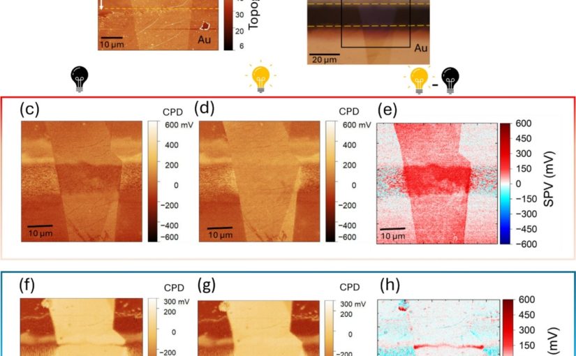
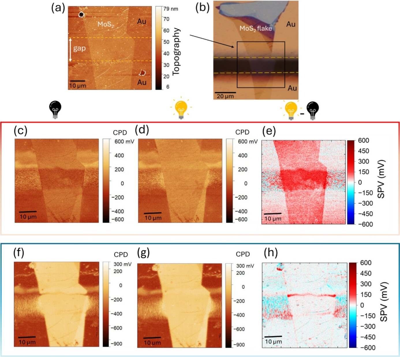
The technique was demonstrated on a silicon photodiode and a molybdenum disulfide (MoS₂) bilayer deposited on a gold substrate. By capturing illuminated and non-illuminated contact potential difference (CPD) measurements along identical scan paths, SM-KPFM produced accurate, drift-free surface photovoltage maps and provided improved insight into nanoscale photovoltaic behavior and charge separation processes in optoelectronic materials.
Kelvin Probe Force Microscopy measurements were performed in sideband mode using a NanoWorld ARROW-EFM AFM probe. The Pt/Ir-coated AFM probe, featuring a resonance frequency of 68 kHz and a spring constant of 2.8 N/m, enabled highly sensitive surface potential mapping with excellent electrical conductivity and measurement stability. The synchronization approach required only triggering the illumination source using the AFM scan direction signal, making the technique readily applicable to existing KPFM workflows without complex hardware modifications.
This work demonstrates how combining an innovative synchronized measurement strategy with a NanoWorld ARROW-EFM AFM probe significantly improves the reliability of operando Kelvin Probe Force Microscopy. The methodology opens new opportunities for investigating nanoscale electronic and optoelectronic devices, photovoltaic materials, and other functional nanostructures where precise surface potential mapping is essential.
Full citation:
Eftekhari, Z.; Ufer, A.; Wurstbauer, U.; Saive, R.
Synchronized modulation Kelvin probe force microscopy for surface photovoltage studies in optoelectronic systems.
MRS Communications 16 (2026), 180–186.
https://doi.org/10.1557/s43579-025-00899-3