
Engineering surfaces with precisely controlled chemical and structural properties is essential for developing advanced biomaterials, tissue engineering platforms, and biofunctional interfaces. In this article, Inga Litzen, Alexander Töpel, Martin Zenke, Antonio Sechi, and Andrij Pich present a versatile strategy for fabricating multifunctional microstructured surfaces by combining microcontact printing with reactive microgel technology.
The authors synthesized poly(N-vinylcaprolactam-co-glycidyl methacrylate) (p(VCL-co-GMA)) microgels that serve as functional colloidal inks for microcontact printing onto glass substrates. This approach enabled the fabrication of stable microgel arrays with well-defined geometries and allowed subsequent post-modification with functional molecules, including fluorescent dyes and cell-adhesive peptide sequences. By further introducing controlled surface-chemical gradients through dip-coating, the researchers demonstrated precise spatial control over surface functionality. Cell culture experiments using NIH-3T3 fibroblasts confirmed that peptide-functionalized microgel patterns significantly influenced cell adhesion and migration, highlighting the potential of this platform for bioengineering and regenerative medicine.
Atomic force microscopy (AFM) was employed to characterize the morphology and surface structure of the synthesized microgels after deposition. High-resolution AFM imaging was performed in tapping mode using a NanoWorld PointProbe® NCH AFM probe. The silicon AFM probe, featuring a resonance frequency of approximately 320 kHz and a spring constant of 42 N/m, enabled detailed visualization of the microgel topography and provided valuable information on particle morphology and surface organization at the nanoscale.
AFM characterization complemented dynamic light scattering and zeta potential measurements by providing direct nanoscale imaging of the surface-coated microgels. The high spatial resolution of the NanoWorld PointProbe® NCH AFM probe allowed the authors to verify the successful formation of uniform microgel structures, supporting the optimization of the microcontact printing process and the subsequent surface functionalization steps.
This article demonstrates how NanoWorld AFM probes contribute to the characterization of advanced polymeric microgel systems used for surface engineering. By combining high-resolution AFM imaging with microcontact printing and post-functionalization strategies, the study provides new opportunities for designing multifunctional surfaces for biomedical, biotechnology, and materials science applications.
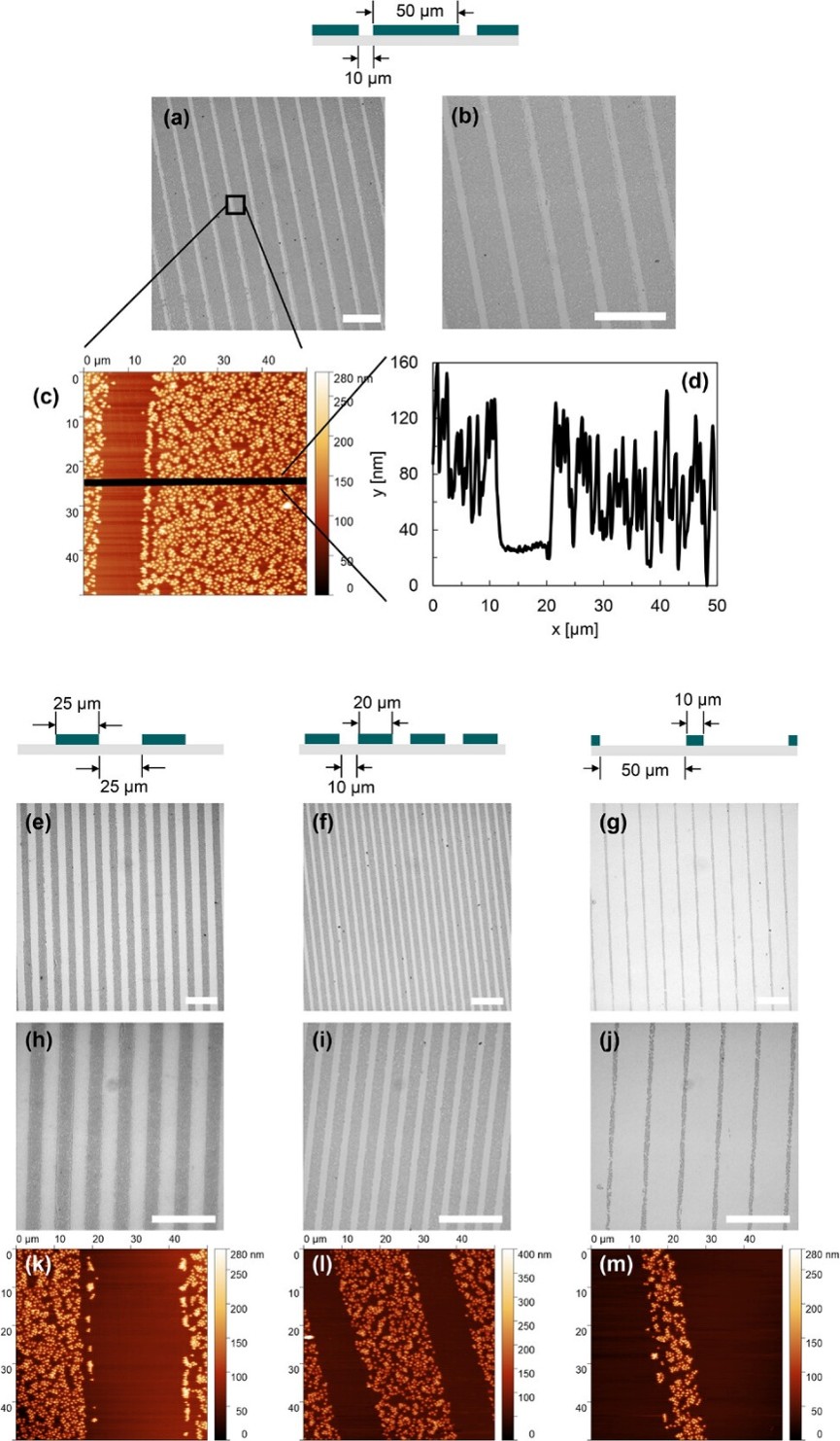
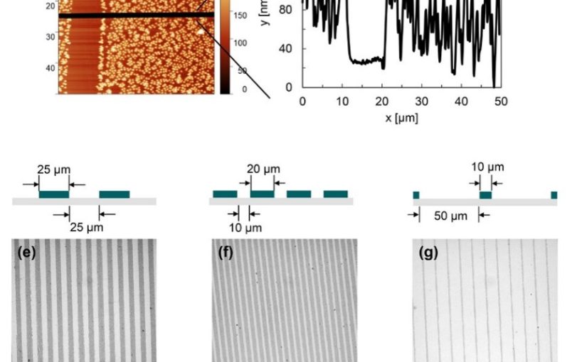
Imaging of surfaces structured with p(VCL-co-10mol%GMA-shell) microgels via microcontact printing. a,b,e-j) Light microscopy images and c,k-m) AFM images were recorded. Additionally, d) shows a height profile generated from the AFM image b). The ideal dimensions of resulting patterns are given in the schemes above. Dark grey areas in light microscopy images represent microgel stripes, lighter areas represent the bare glass. Scale bars in light microscopy images: 100 µm.
Full citation:
Litzen, I.; Töpel, A.; Zenke, M.; Sechi, A.; Pich, A.
Multifunctional Microstructured Surfaces by Microcontact Printing of Reactive Microgels.
Advanced Functional Materials 36(15), e16135 (2026).
https://doi.org/10.1002/adfm.202516135
Creative Commons license: CC BY 4.0