We have a month with “R” again and the shellfish season has started in the Northern Hemisphere. So we’d like to share the Nature Communications article by Petrone et. al “Mussel adhesion is dictated by time-regulated secretion and molecular conformation of mussel adhesive proteins” with you.
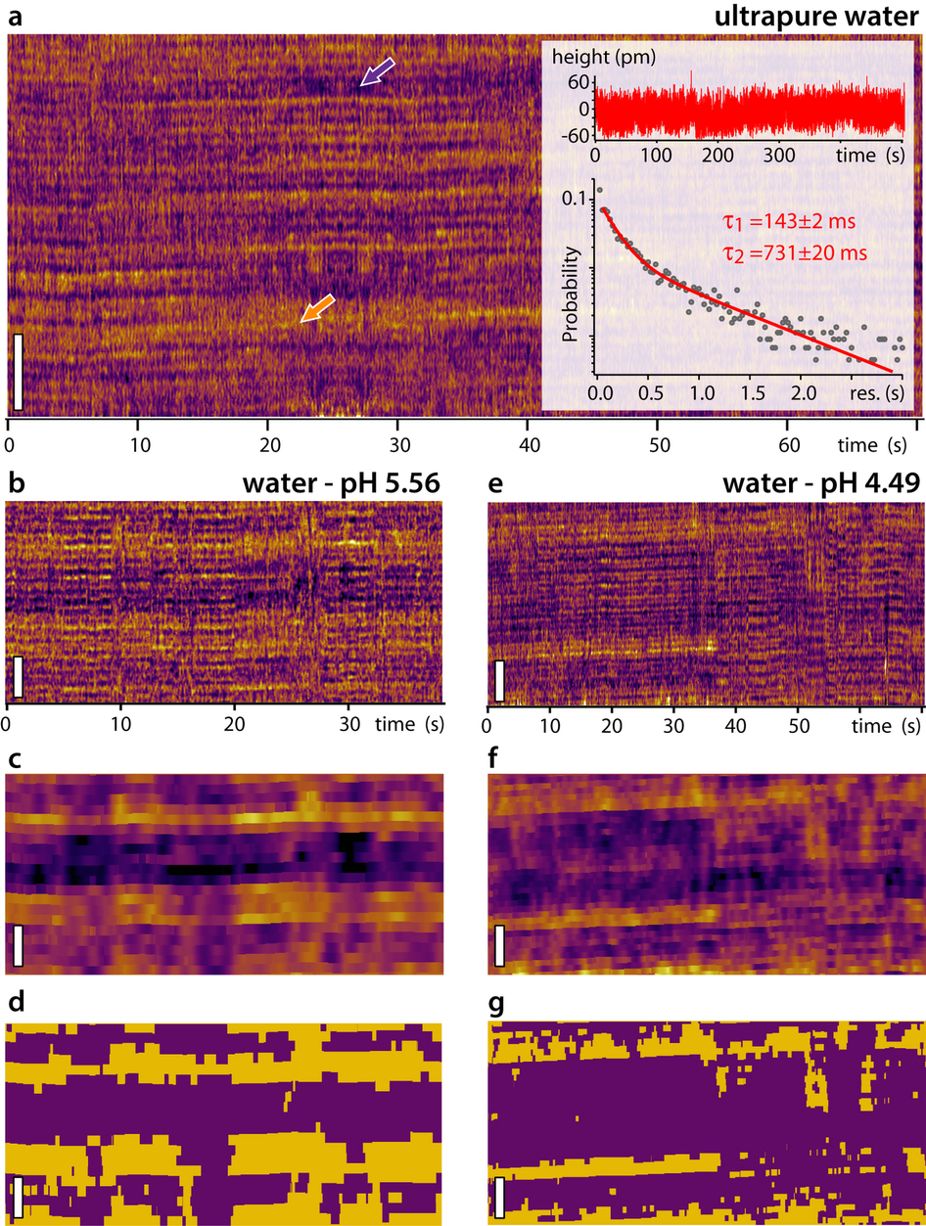
A NanoWorld Pointprobe® NCSTR AFM probe was used for the AFM images in this paper. This AFM probe is designed to give extra stability and accuracy during soft tapping mode imaging in order to produce higher quality AFM images while minimizing sample damage.

secretion and molecular conformation of mussel adhesive proteins”:
Atomic Force Microscopy (AFM) of mussel adhesive proteins on mica. AFM images of dry Pvfp-3α and Pvfp-5β adsorbed from 0.02 mg ml-1 solution in 5% acetic acid and 0.25 MO3 on mica. After 20 min adsorption, the mica surfaces were washed with protein -free buffer, and the AFM images show the homogenous distribution of the resulting adsorbed proteins. The height profiles for both proteins are shown in the graphs below, corresponding to the dotted red and blue lines in the respective AFM images (see black arrows).
Luigi Petrone, Akshita Kumar, Clarinda N. Sutanto, Navinkumar J. Patil, Srinivasaraghavan Kannan, Alagappan Palaniappan, Shahrouz Amini, Bruno Zappone, Chandra Verma, Ali Miserez
Mussel adhesion is dictated by time-regulated secretion and molecular conformation of mussel adhesive proteins
Nature Communications volume 6, Article number: 8737 (2015)
DOI https://doi.org/10.1038/ncomms9737
Please follow this external link for the full article: https://rdcu.be/5vcI
The article by Petrone, L.et al. “Mussel adhesion is dictated by time-regulated secretion and molecular conformation of mussel adhesive proteins” is licensed under a Creative Commons Attribution 4.0 International License. The images or other third party material in this article are included in the article’s Creative Commons license, unless indicated otherwise in the credit line; if the material is not included under the Creative Commons license, users will need to obtain permission from the license holder to reproduce the material. To view a copy of this license, visit http://creativecommons.org/licenses/by/4.0/