Kelvin Probe Force Microscopy ( KPFM ) is a scanning probe microscopy technique. It is a combination of the Kelvin probe and of Atomic Force Microscopy methods. The technique consists in evaluating the difference in work function between two conducting materials, by using a nanometer scale tip ( the “KPFMtip”), and placing it close to the material to be characterised, where a difference in work function leads to an electrostatic force developing between the two, which is translated as an oscillation of the tip’s cantilever. A bia sapplied via an external circuit is varied until the force and hence the electrostatic field between sample and KPFM tip is cancelled.*
In the
article “KPFM surface photovoltage measurement and numerical simulation”
Clément Marchat, James P. Connolly, Jean-Paul Kleider, José Alvarez, Lejo J.
Koduvelikulathu and Jean Baptiste Puel present a method for the analysis of
Kelvin probe force microscopy (KPFM) characterization of semiconductor devices.
It enables evaluation of the influence of defective surface layers. The model
is validated by analysing experimental KPFM measurements on crystalline silicon
samples of contact potential difference (VCPD) in the dark and under
illumination, and hence the surface photovoltage (SPV). It is shown that the
model phenomenologically explains the observed KPFM measurements. It reproduces
the magnitude of SPV characterization as a function of incident light power in
terms of a defect density assuming Gaussian defect distribution in the
semiconductor bandgap. This allows an estimation of defect densities in surface
layers of semiconductors and therefore increased exploitation of KPFM data.*
The KPFM measurements were performed using NanoWorld ARROW-EFM conductive AFM tips with a PtIr coating.
The tip work function didn’t require calibration because only SPV measurement were performed and studied. Measurements were performed in the KPFM amplitude modulation (AM)mode rather than the frequency modulation (FM) one. The AM mode was chosen because lateral resolution was not a problem on the homogeneous bulk samples studied, allowing focus on the superior surface potential resolution that can be achieved with the AM mode.*
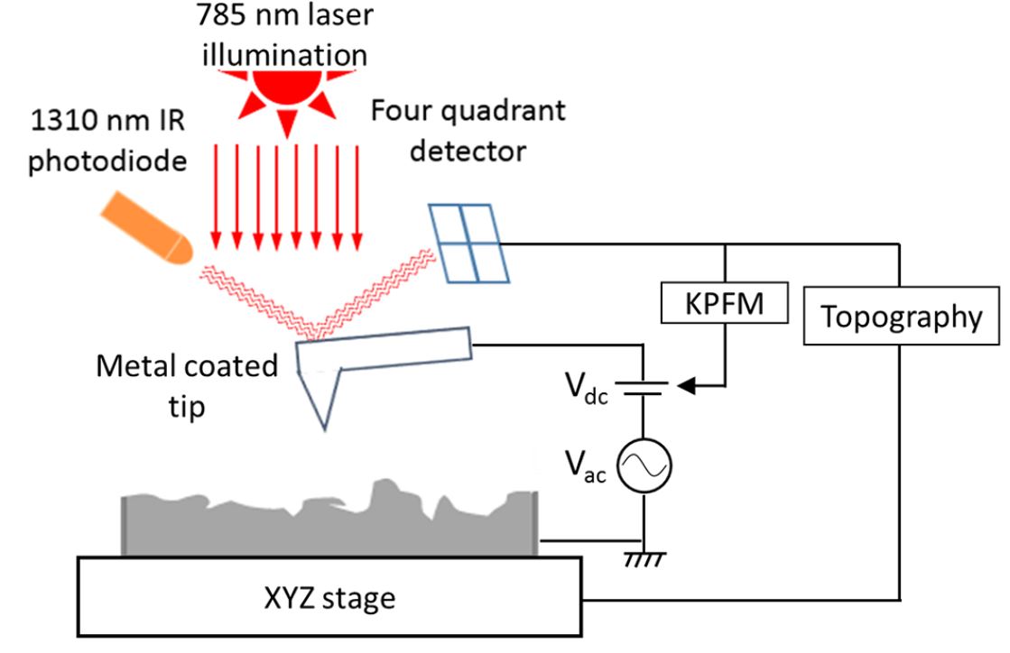
Kelvin probe force microscopy setup schematic. The conducting cantilever carrying the KPFM tip is scanned over a surface while AC + DC potential is applied. The AC signal is a sinusoid whose frequency matches the mechanical resonance of the cantilever. The four-quadrant detector provides feedback in order to minimise cantilever oscillation by varying the DC signal thereby yielding the sample work function compared to the tip one.
*Clément Marchat, James P. Connolly, Jean-Paul Kleider, José Alvarez, Lejo J. Koduvelikulathu and Jean Baptiste Puel
KPFM surface photovoltage measurement and numerical simulation
EPJ Photovoltaics10, 3 (2019)
DOI: https://doi.org/10.1051/epjpv/2019002
Please follow this external link to read the full article: https://www.epj-pv.org/articles/epjpv/abs/2019/01/pv180014/pv180014.html
Open Access The article “KPFM surface photovoltage measurement and numerical simulation “ by Clément Marchat, James P. Connolly, Jean-Paul Kleider, José Alvarez, Lejo J. Koduvelikulathu and Jean Baptiste Puel is licensed under a Creative Commons Attribution 4.0 International License, which permits use, sharing, adaptation, distribution and reproduction in any medium or format, as long as you give appropriate credit to the original author(s) and the source, provide a link to the Creative Commons license, and indicate if changes were made. The images or other third party material in this article are included in the article’s Creative Commons license, unless indicated otherwise in a credit line to the material. If material is not included in the article’s Creative Commons license and your intended use is not permitted by statutory regulation or exceeds the permitted use, you will need to obtain permission directly from the copyright holder. To view a copy of this license, visit http://creativecommons.org/licenses/by/4.0/.