The current pandemic is not the only health threat worldwide. Another worry is the increasing antibiotic resistance which increases the fear to run out of effective antibiotics.
This is one of the reasons why antimicrobial peptides (AMPs) are gaining more and more interest.
The lipopeptide Daptomycin ( DAP ) has been therapeutically used as a last resort antibiotic against multidrug-resistant enterococci and staphylococci in the past. Unfortunately, some strains have become resistant to Dap in recent years. There still is a knowledge-gap on the details of Dap activity. It is therefore important to understand the structure-activity relationships of AMPs on membranes in order to develop more antibiotics of this type as a countermeasure to the spread of resistance.*
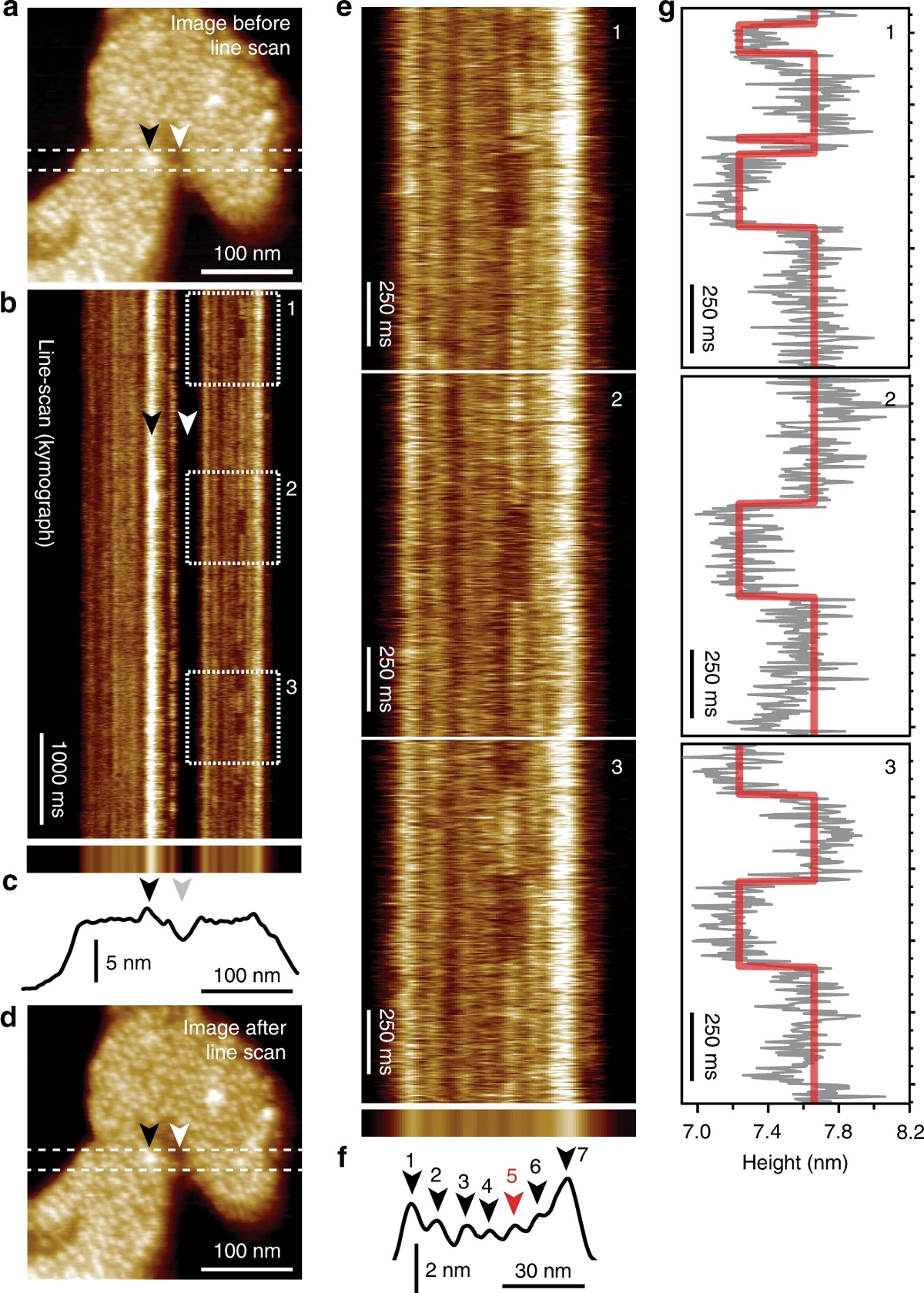
High Speed Atomic Force Microscopy ( HS-AFM ) makes it possible to observe dynamic biological processes on a molecular level.
In the article “High-speed atomic force microscopy highlights new molecular mechanism of daptomycin action” Francesca Zuttion, Adai Colom, Stefan Matile, Denes Farago, Frédérique Pompeo, Janos Kokavecz, Anne Galinier, James Sturgis and Ignacio Casuso describe how, by using the possibilities offered by high speed atomic force microscopy, they were able to confirm some up until now hypothetical models and additionally detected some previously unknown molecular mechanisms. *
The HS-AFM imaging made it possible for the authors to observe the development of the dynamics of interaction at the molecular-level over several hours. *
They investigated the lipopeptide Daptomycin under infection-like conditions and could confirm Dap oligomerization and the existence of half pores. *
They also mimicked bacterial resistance conditions by increasing the CL-content in the membrane. *
By correlating the results of other research techniques such as FRET, SANS, NMR, CD or electrophysiology techniques with the results they achieved with high speed atomic force microscopy F. Zuttion et al. were able to confirm several, previously, hypothetical models, and detect several unknown molecular mechanisms. *
It is to be hoped that the possibilities offered by HS-AFM imaging will stimulate new models and insight on the structure-activity relationship of membrane-interacting molecules and also open up the possiblity to increase the throughput of screening of molecular candidates considerably. *
NanoWorld USC ( Ultra-Short AFM Cantilevers) of the USC-F1.2-k0.15 type, which are specially designed for the use in high speed atomic force microscopy, were used for the HS-AFM imaging described in the article cited below. These AFM probes have a typical resonance frequency of 1200 kHz and have a wear resistant AFM tip made from high density carbon.

Intermediate stages a A new structure appeared: dimples, zones of thinner membrane thickness, whose diameter was in the range 7 ± 2 nm. Most dimples diffuse, but some remained static (colour scale: 3 nm). Movie details: frame rate 97 ms; zoom of a full image of 150 nm × 90 nm and 256 × 160 pixels. b The dimple diffusion consisted of swinging trajectories, implying membrane-mediated dimple-dimple attraction (colour scale: 3 nm). b, right, Energy profile of the interaction of the dimples obtained derived from 120 centre-to-centre distance measurements that contains as the oligomers two energy minima. Movie details: frame rate 83 ms; full image of 150 nm × 150 nm and 256 × 256 pixels. c In some membrane zones, clusters of dimples, reminiscent of cubic phases, developed (colour scale: 4 nm). Movie details: frame rate 74 ms; full image of 90 nm × 60 nm and 256 × 160 pixels. d The clusters of dimples were moderately dynamical in time, with moderate internal rearrangements (colour scale: 4 nm). Movie details: frame rate 74 ms; full image of 25 nm × 16 nm and 256 × 160 pixels. e The other deformation found was elongated-humps on top of the POPG membrane. e, left, An elongated-hump in the proximity of a cluster of dimples (colour scale: 4 nm). e, right, A close-up and a profile of an elongated-hump. Additional images of elongated-humps on Supplementary Fig. 1. Movie details: frame rate 479 ms; zoom of full image of 250 nm × 200 nm and 300 × 256 pixels. f It was observed that the dimples and the elongated-humps fused and gave yield to pores of toroidal structure where a protruding ring surrounds the pore (colour scale: 4 nm). Movie details: frame rate 74 ms; full image of 40 nm × 40 nm and 256 × 160 pixels.
*Francesca Zuttion, Adai Colom, Stefan Matile, Denes Farago, Frédérique Pompeo, Janos Kokavecz, Anne Galinier, James Sturgis and Ignacio Casuso
High-speed atomic force microscopy highlights new molecular mechanism of daptomycin action
Nature Communications volume 11, Article number: 6312 (2020)
DOI: https://doi.org/10.1038/s41467-020-19710-z
Please follow this external link to read the full article: https://rdcu.be/ciaW2
Open Access : The article “High-speed atomic force microscopy highlights new molecular mechanism of daptomycin action” by Francesca Zuttion, Adai Colom, Stefan Matile, Denes Farago, Frédérique Pompeo, Janos Kokavecz, Anne Galinier, James Sturgis and Ignacio Casuso is licensed under a Creative Commons Attribution 4.0 International License, which permits use, sharing, adaptation, distribution and reproduction in any medium or format, as long as you give appropriate credit to the original author(s) and the source, provide a link to the Creative Commons license, and indicate if changes were made. The images or other third party material in this article are included in the article’s Creative Commons license, unless indicated otherwise in a credit line to the material. If material is not included in the article’s Creative Commons license and your intended use is not permitted by statutory regulation or exceeds the permitted use, you will need to obtain permission directly from the copyright holder. To view a copy of this license, visit https://creativecommons.org/licenses/by/4.0/.