In the article “Multiswitchable photoacid–hydroxyflavylium–polyelectrolyte nano-assemblies” Alexander Zika and Franziska Gröhn describe the development of a novel reversible multi-switchable system consisting of a cationic polyelectrolyte, a hydroxyflavylium molecule (Flavy), and a photoacid.*
Ternary assemblies with sizes in the hundred-to-few hundred nanometers range in aqueous solution exhibit a multi-addressable size and shape.*
The concept exploits the unique property of the photoacid to form a more highly charged molecule and to switch the Flavy molecule in the same step when excited by light irradiation.*
Due to the network of possible reactions of Flavy, self-assembly can be accessed and triggered in a number of ways.*
While their study focused on the first proof of concept and the relation of molecular and nanoscale switching, a deeper understanding of the molecular binding effects may be considered in future studies.*
The type of the photoacid-based assembly presented in the article bears potential, for example, for delivery where the assembly property changes may provide a desirable transformable platform for tunable and smart transport.*
Figure 6 from “Multiswitchable photoacid–hydroxyflavylium–polyelectrolyte nano-assemblies” by Alexander Zika and Franziska Gröhn: Comparison of cycle I and cycle II in AFM.
*Alexander Zika and Franziska Gröhn Multiswitchable photoacid–hydroxyflavylium–polyelectrolyte nano-assemblies Beilstein J. Org. Chem. 2021, 17, 166–185. DOI: https://doi.org/10.3762/bjoc.17.17
Open Access : The article “Multiswitchable photoacid–hydroxyflavylium–polyelectrolyte nano-assemblies” by Alexander Zika and Franziska Gröhn is licensed under a Creative Commons Attribution 4.0 International License, which permits use, sharing, adaptation, distribution and reproduction in any medium or format, as long as you give appropriate credit to the original author(s) and the source, provide a link to the Creative Commons license, and indicate if changes were made. The images or other third party material in this article are included in the article’s Creative Commons license, unless indicated otherwise in a credit line to the material. If material is not included in the article’s Creative Commons license and your intended use is not permitted by statutory regulation or exceeds the permitted use, you will need to obtain permission directly from the copyright holder. To view a copy of this license, visit https://creativecommons.org/licenses/by/4.0/.
Excitatory amino acid transporters (EAATs) are important in many physiological processes and crucial for the removal of excitatory amino acids from the synaptic cleft.*
In the article “Millisecond dynamics of an unlabeled amino acid transporter “ Tina R. Matin, George R. Heath, Gerard H. M. Huysmans, Olga Boudker and Simon Scheuring develop and apply high-speed atomic force microscopy line-scanning (HS-AFM-LS) combined with automated state assignment and transition analysis for the determination of transport dynamics of unlabeled membrane-reconstituted GltPh, a prokaryotic EAAT homologue, with millisecond temporal resolution.*
Among the bulk and single-molecule techniques, high-speed atomic force microscopy ( HS-AFM ) stands out with its ability to provide real-time structural and dynamical information of single molecules. HS-AFM images label-free molecules under close-to-physiological conditions with ~0.1 nm vertical and ~1 nm lateral imaging resolution. Furthermore, HS-AFM has typically ~100 ms temporal resolution, giving access to structure–dynamics relationship of proteins, though the achievable imaging speed depends on sample characteristics like scan size and surface corrugation.
Recently in a quest to achieve higher temporal resolutions, the authors of the cited article used HS-AFM line scanning (HS-AFM-LS) for the analysis of single-protein dynamics. *
Line scanning, using a conventional AFM, has been used to study protein–protein interactions earlier. In HS-AFM-LS, the slow-scan axis (y-direction) is disabled. Therefore, instead of imaging an x/y-area, the scientists scan over one horizontal x-line several hundreds to thousands of times per second, thus reaching millisecond temporal resolution. The topographical readouts of this line are stacked one after another, resulting in kymographs of the dynamical behavior of the molecules. Therefore, HS-AFM-LS has between 2 and 3 orders of magnitude higher temporal resolution than HS-AFM imaging and should allow the detection of fast transporter dynamics and possible intermediate states that have so far escaped kinetic characterization. *
All AFM images presented in this study were taken using a HS-AFM operated in amplitude modulation mode (with typical free and setpoint amplitudes, Afree = 1.0 nm and Aset = 0.9 nm, respectively using optimized scan and feedback parameters. NanoWorld Ultra-Short Cantilevers ( NanoWorld’s AFM probe series especially dedicated for High Speed Scanning) of the USC-F1.2-k0.15 type were used. In the presented experiments, four different buffer conditions were used. *
As the authors state in their article they find that GltPh transporters can operate much faster than previously reported, with state dwell-times in the 50 ms range, and report the kinetics of an intermediate transport state with height between the outward- and inward-facing states. Transport domains stochastically probe transmembrane motion, and reversible unsuccessful excursions to the intermediate state occur. The presented approach and analysis methodology are generally applicable to study transporter kinetics at system-relevant temporal resolution.*
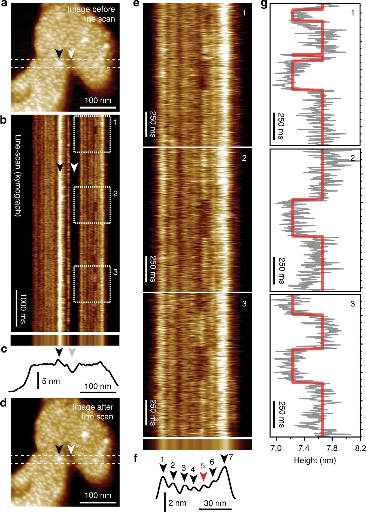
Figure 2 from “Millisecond dynamics of an unlabeled amino acid transporter” by Tina R. Matin et al. HS-AFM line scanning (HS-AFM-LS): millisecond temporal resolution of unlabeled transporter dynamics.: a HS-AFM image of a membrane packed with GltPh exposing the extracellular face before HS-AFM-LS (apo condition: 20 mM Tris-HCl, pH7.5, 150 mM KCl). Dashed lines indicate the position of the central scan line where subsequent HS-AFM-LS is performed. b Six seconds of a HS-AFM-LS kymograph with 3.3 ms line acquisition speed. Each transporter domain appears as a vertical line. c Projection (top) and height profile (bottom) of b. d HS-AFM image after HS-AFM-LS. The lateral position of recognizable features in a–d are indicated by arrowheads. e One second high-magnification views of dashed regions 1, 2, and 3 in b. Transport domain excursions to the inward-facing state appear as dark dwells along the vertical time axis. f Projection (top) and height profile (bottom) of e. Arrowheads indicate the position of the seven protomers in the kymograph (red: active protomer #5). g Height/time traces (gray) and state fits (red) of the active domain (protomer #5) in e. This figure is representative of the experimental sequence for the >50 replicates analyzed in this work.
*Tina R. Matin, George R. Heath, Gerard H. M. Huysmans, Olga Boudker and Simon Scheuring Millisecond dynamics of an unlabeled amino acid transporter Nature Communications volume 11, Article number: 5016 (2020) DOI: https://doi.org/10.1038/s41467-020-18811-z
Open Access : The article “Millisecond dynamics of an unlabeled amino acid transporter” by Tina R. Matin, George R. Heath, Gerard H. M. Huysmans, Olga Boudker and Simon Scheuring is licensed under a Creative Commons Attribution 4.0 International License, which permits use, sharing, adaptation, distribution and reproduction in any medium or format, as long as you give appropriate credit to the original author(s) and the source, provide a link to the Creative Commons license, and indicate if changes were made. The images or other third party material in this article are included in the article’s Creative Commons license, unless indicated otherwise in a credit line to the material. If material is not included in the article’s Creative Commons license and your intended use is not permitted by statutory regulation or exceeds the permitted use, you will need to obtain permission directly from the copyright holder. To view a copy of this license, visit https://creativecommons.org/licenses/by/4.0/.
Lipid membranes play a key role in living systems by providing a structural barrier that separates cellular compartments. Bilayer fluidity in the lateral plane is a key property of lipid membranes, that allows the membrane to have sufficient flexibility to accommodate dynamic stresses, shape changes and rearrangements accompanying the cellular lifecycle.*
In the article “Carbon nanotube porin diffusion in mixed composition supported lipid bilayers” Kylee Sullivan, Yuliang Zhang, Joseph Lopez, Mary Lowe and Aleksandr Noy describe how they used high-speed atomic force microscopy (HS-AFM) and all-atom molecular dynamics (MD) simulations to study the behavior of CNTPs in a mixed lipid membrane consisting of DOPC lipid with a variable percentage of DMPC lipid added to it. HS-AFM data reveal that the CNTPs undergo diffusive motion in the bilayer plane.*
Motion trajectories extracted from the HS-AFM movies indicate that CNTPs exhibit diffusion coefficient values broadly similar to values reported for membrane proteins in supported lipid bilayers. The data also indicate that increasing the percentage of DMPC leads to a marked slowing of CNTP diffusion. MD simulations reveal a CNTP-lipid assembly that diffuses in the membrane and show trends that are consistent with the experimental observations. *
The above-mentioned study confirms that CNTPs mimic the major features of the diffusive movement of biological pores in lipid membranes and shows how the increase in bilayer viscosity leads to a corresponding slowdown in protein motion. It should be possible to extend this approach to studies of other membrane protein dynamics in supported lipid bilayers. The authors note that those studies, however, will need to be mindful of the challenge of unambiguous visualization of the membrane components, especially in systems that incorporate smaller proteins, such as antimicrobial peptides. Another challenge that could complicate these studies would be microscopic phase separation of the lipid matrix that could lead to complicated pore dynamics in the membrane. *
NanoWorld Ultra-Short AFM cantilevers with high-density carbon/diamond-like carbon (HDC/DLC) AFM tips of the USC-F1.2-k0.15 type were used for the high-speed atomic force microscopy described in the article. *
Figure 2 a and b from “Carbon nanotube porin diffusion in mixed composition supported lipid bilayers” by Kylee Sullivan et al.:
CNTP motion in supported lipid bilayers. (a) Representative frames (with times in seconds indicated on each image) from an HS-AFM movie showing a CNTP diffusing in a supported lipid bilayer with 80:20 DOPC-DMPC ratio (see also Supplementary Movie 2). (b) A representative trajectory for CNTP diffusion in the bilayer. The time step between each datapoint is 0.5 s. Please refer tothe full article cited below for thefull figure.
*Kylee Sullivan, Yuliang Zhang, Joseph Lopez, Mary Lowe and Aleksandr Noy Carbon nanotube porin diffusion in mixed composition supported lipid bilayers Nature Scientific Reports volume 10, Article number: 11908 (2020) DOI: https://doi.org/10.1038/s41598-020-68059-2
Open Access : The article “Carbon nanotube porin diffusion in mixed composition supported lipid bilayers” by Kylee Sullivan, Yuliang Zhang, Joseph Lopez, Mary Lowe and Aleksandr Noy is licensed under a Creative Commons Attribution 4.0 International License, which permits use, sharing, adaptation, distribution and reproduction in any medium or format, as long as you give appropriate credit to the original author(s) and the source, provide a link to the Creative Commons license, and indicate if changes were made. The images or other third party material in this article are included in the article’s Creative Commons license, unless indicated otherwise in a credit line to the material. If material is not included in the article’s Creative Commons license and your intended use is not permitted by statutory regulation or exceeds the permitted use, you will need to obtain permission directly from the copyright holder. To view a copy of this license, visit https://creativecommons.org/licenses/by/4.0/.