The free energy landscape of retroviral integration

Retroviral integration, the process of covalently inserting viral DNA into the host genome, is a point of no return in the replication cycle. Yet, strand transfer is intrinsically iso-energetic and it is not clear how efficient integration can be achieved.*

In the article “The free energy landscape of retroviral integration” published in Nature Communications Willem Vanderlinden, Tine Brouns, Philipp U. Walker, Pauline J. Kolbeck, Lukas F. Milles, Wolfgang Ott, Philipp C. Nickels, Zeger Debyser and Jan Lipfert use biochemical assays, atomic force microscopy (AFM), and multiplexed single-molecule magnetic tweezers (MT) to study tetrameric prototype foamy virus (PFV) strand-transfer dynamics.*

Their finding that PFV intasomes employ auxiliary-binding sites for modulating the barriers to integration raises the question how the topology of higher-order intasomes governs integration of pathogenic retroviruses, most notably HIV. The single-molecule assays developed in this work are expected to be particularly useful to further unravel the complexity of this important class of molecular machines.*

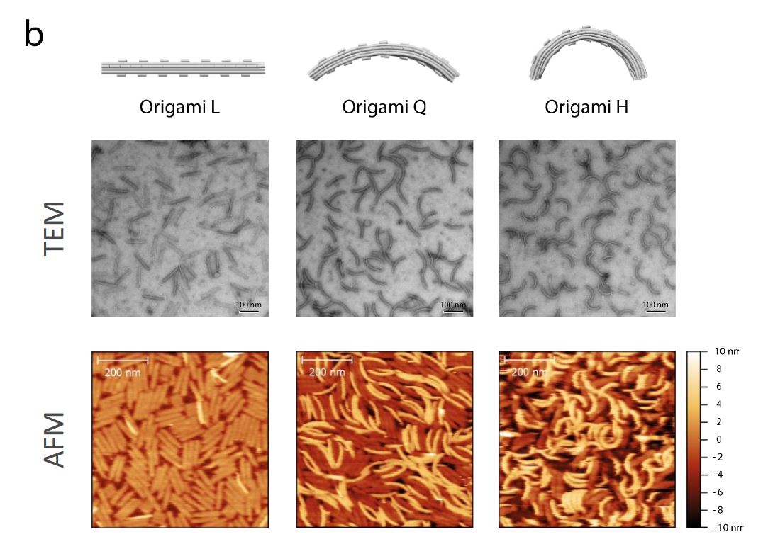
The AFM images were recorded in amplitude modulation mode under ambient conditions and by using NanoWorld high resolution SuperSharpSiliconSSS-NCH cantilevers ( resonance frequency ≈300 kHz; typical end-radius 2 nm; half-cone angle <10 deg). Typical scans were recorded at 1–3 Hz line frequency, with optimized feedback parameters and at 512 × 512 pixels.*

Figure 2 e, f and g from “The free energy landscape of retroviral integration” by Willem Vanderlinden et al. 
(please refer to the full article for the complete figure 2  https://rdcu.be/b0R63 ) :
  e Atomic Force Microscopy image of intasomes incubated briefly (2 min) with supercoiled plasmid DNA, depicting a branched complex as found in ~50% of early complexes.
  f  Atomic Force Microscopy image of a bridging complex that dominates (~80%) the population of complexes at longer (>45 min) incubation. 
 g  Atomic Force Microscopy image of a gel-purified STC
Figure 2 e, f and g from “The free energy landscape of retroviral integration” by Willem Vanderlinden et al.
(please refer to the full article for the complete figure 2 https://rdcu.be/b0R63 ) :
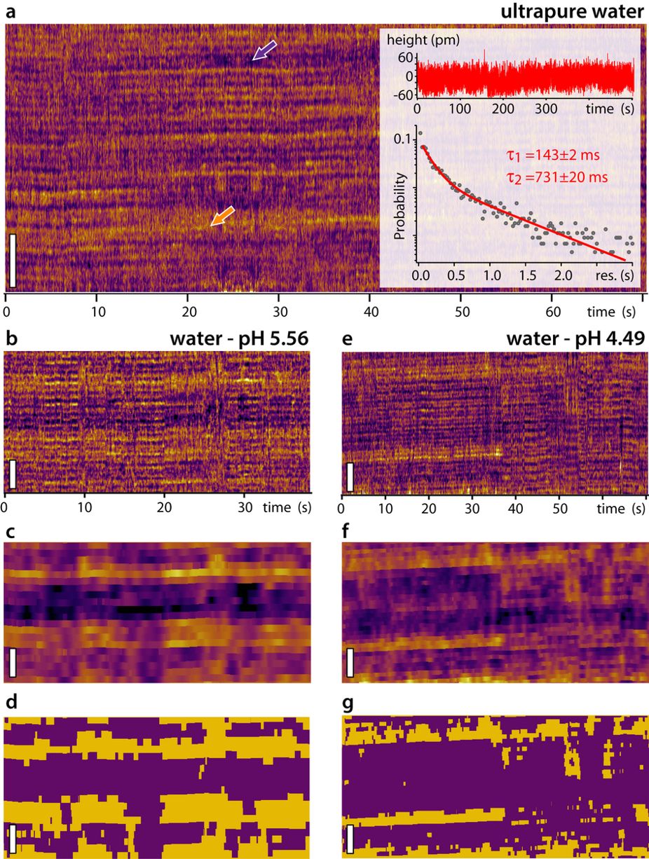
 e AFM image of intasomes incubated briefly (2 min) with supercoiled plasmid DNA, depicting a branched complex as found in ~50% of early complexes.
 f AFM image of a bridging complex that dominates (~80%) the population of complexes at longer (>45 min) incubation.
g AFM image of a gel-purified STC

*Willem Vanderlinden, Tine Brouns, Philipp U. Walker, Pauline J. Kolbeck, Lukas F. Milles, Wolfgang Ott, Philipp C. Nickels, Zeger Debyser, Jan Lipfert
The free energy landscape of retroviral integration
Nature Communications volume 10, Article number: 4738 (2019)
DOI: https://doi.org/10.1038/s41467-019-12649-w

Please follow this external link to read the full article: https://rdcu.be/b0R63

Open Access The article “The free energy landscape of retroviral integration“ by Willem Vanderlinden, Tine Brouns, Philipp U. Walker, Pauline J. Kolbeck, Lukas F. Milles, Wolfgang Ott, Philipp C. Nickels, Zeger Debyser and Jan Lipfert is licensed under a Creative Commons Attribution 4.0 International License, which permits use, sharing, adaptation, distribution and reproduction in any medium or format, as long as you give appropriate credit to the original author(s) and the source, provide a link to the Creative Commons license, and indicate if changes were made. The images or other third party material in this article are included in the article’s Creative Commons license, unless indicated otherwise in a credit line to the material. If material is not included in the article’s Creative Commons license and your intended use is not permitted by statutory regulation or exceeds the permitted use, you will need to obtain permission directly from the copyright holder. To view a copy of this license, visit http://creativecommons.org/licenses/by/4.0/.

Membrane sculpting by curved DNA origami scaffolds

In the article “Membrane sculpting by curved DNA origami scaffolds” the authors show that “dependent on curvature, membrane affinity and surface density, DNA origami coats can indeed reproduce the activity of membrane-sculpting proteins such as BAR, suggesting exciting perspectives for using them in bottom-up approaches towards minimal biomimetic cellular machineries.”*

The AFM images for this article were taken in high-speed AC mode using NanoWorld Ultra-Short Cantilevers of the USC-F0.3-k0.3 type.

Supplementary Figure 5 b from Membrane sculpting by curved DNA origami scaffolds: Characterization of folded DNA origami nanoscaffolds. ( a ) Assembly of the folded bare origami structures L, Q, H was initially assessed via agarose gel (2%) electrophoresis analysis. Lanes containing marker DNA ladder (1kb) and M13 single - stranded p7249 sc affold (Sc) were also included. ( b ) Structure of folded bare origami L, Q and H was further validated using negative - stain transmission electron microscopy (TEM ; scale bar s : 100 nm ) and atomic force microscopy (AFM ; scale bar s : 200nm ).
Supplementary Figure 5 b from “Membrane sculpting by curved DNA origami scaffolds”:
Characterization of folded DNA origami nanoscaffolds.
b) Structure of folded bare origami L, Q and H was further validated using negative-stain transmission electron microscopy (TEM; scale bars: 100 nm) and atomic force microscopy (AFM; scale bars: 200nm).

*Henri G. Franquelim, Alena Khmelinskaia, Jean-Philippe Sobczak, Hendrik Dietz, Petra Schwille
Membrane sculpting by curved DNA origami scaffolds
Nature Communicationsvolume 9, Article number: 811 (2018)
DOI: https://doi.org/10.1038/s41467-018-03198-9

Please follow this link to the full article: https://rdcu.be/8zZi

Open Access: The article “Membrane sculpting by curved DNA origami scaffolds” by Franquelim et. al is licensed under a Creative Commons Attribution 4.0 International License, which permits use, sharing, adaptation, distribution and reproduction in any medium or format, as long as you give appropriate credit to the original author(s) and the source, provide a link to the Creative Commons license, and indicate if changes were made. The images or other third party material in this article are included in the article’s Creative Commons license, unless indicated otherwise in a credit line to the material. If material is not included in the article’s Creative Commons license and your intended use is not permitted by statutory regulation or exceeds the permitted use, you will need to obtain permission directly from the copyright holder. To view a copy of this license, visit http://creativecommons.org/licenses/by/4.0/.

Direct observation of the dynamics of single metal ions at the interface with solids in aqueous solutions

For the AFM measurements in the article “Direct observation of the dynamics of single metal ions at the interface with solids in aqueous solutions” by Ricci, M. et al. a NanoWorld Arrow-UHFAuD AFM probe was used. Congratulations to the authors!

Figure 3 from: "Ricci, M. et al. Direct observation of the dynamics of single metal ions at the interface with solids in aqueous solutions."
Figure 3 from: “Ricci, M. et al. Direct observation of the dynamics of single metal ions at the interface with solids in aqueous solutions.“: Kinetic experiments conducted in pure water (a) show mainly two levels (arrows) when compared to Fig. 2a. Height variations are less pronounced than in RbCl solution and analysis of the surface dynamics (inset) reveals slower timescales with a relatively strong dependence on the choice of threshold. The profile shown in the inset is taken after site averaging (see e.g. Fig. 2d), hence the small height variations. More reliable results were obtained for lower threshold values (here −20 pm, see Supplementary Fig. S10). The overall ratio between the two levels visible in (a) can be changed by adjusting the pH of the water with HCl (b–g), suggesting the higher level to be related to hydration water and the lower level to reflect adsorption of H3O+, as detected by the AFM tip. For each of the pH value studied, the raw kinetic experiments (b,e) are site-averaged (c,f) as in Fig. 2d to remove the mica corrugation and imaging noise. The height distribution of the site-averaged data is then binarised automatically (d,g) depending on whether the surface height is higher or lower than the average between the surface’s highest and lowest points. The fraction of surface interpreted as covered with H3O+ (purple in d and g) changes from 55 ± 3% to 75 ± 2%. (b,e) were acquired with a same tip. The mica samples have been rinsed with the imaging solution after being cleaved and the presence of K+ ions on the surface can be neglected (concentration <10 nM). The scale bar is 3 nm in all experiments.

Abstract:
The dynamics of ions adsorbed at the surface of immersed charged solids plays a central role in countless natural and industrial processes such as crystal growth, heterogeneous catalysis, electrochemistry, or biological function. Electrokinetic measurements typically distinguish between a so-called Stern layer of ions and water molecules directly adsorbed on to the solid’s surface, and a diffuse layer of ions further away from the surface. Dynamics within the Stern layer remain poorly understood, largely owing to a lack of in-situ atomic-level insights. Here we follow the dynamics of single Rb+ and H3O+ ions at the surface of mica in water using high-resolution atomic force microscopy with 25 ms resolution. Our results suggest that single hydrated Rb+ions reside τ1 = 104 ± 5 ms at a given location, but this is dependent on the hydration state of the surface which evolves on a slower timescale of τ2 = 610 ± 30 ms depending on H3O+ adsorption. Increasing the liquid’s temperature from 5 °C to 65 °C predictably decreases the apparent glassiness of the interfacial water, but no clear effect on the ions’ dynamics was observed, indicating a diffusion-dominated process. These timescales are remarkably slow for individual monovalent ions and could have important implications for interfacial processes in electrolytes.

Maria Ricci, William Trewby, Clodomiro Cafolla, Kislon Voïtchovsky
Direct observation of the dynamics of single metal ions at the interface with solids in aqueous solutions
Nature Scientific Reports volume 7, Article number: 43234 (2017)
doi: https://doi.org/10.1038/srep43234

Please follow this external link for the full article: https://rdcu.be/4QVb

This article “Direct observation of the dynamics of sigle metal ions at the interface with solids in aqueous solutions” by Ricci, M. et al. is licensed under a Creative Commons Attribution 4.0 International License. The images or other third party material in this article are included in the article’s Creative Commons license, unless indicated otherwise in the credit line; if the material is not included under the Creative Commons license, users will need to obtain permission from the license holder to reproduce the material. To view a copy of this license, visit http://creativecommons.org/licenses/by/4.0/