For the AFM measurements in the article “Direct observation of the dynamics of single metal ions at the interface with solids in aqueous solutions” by Ricci, M. et al. a NanoWorld Arrow-UHFAuD AFM probe was used. Congratulations to the authors!
Abstract:
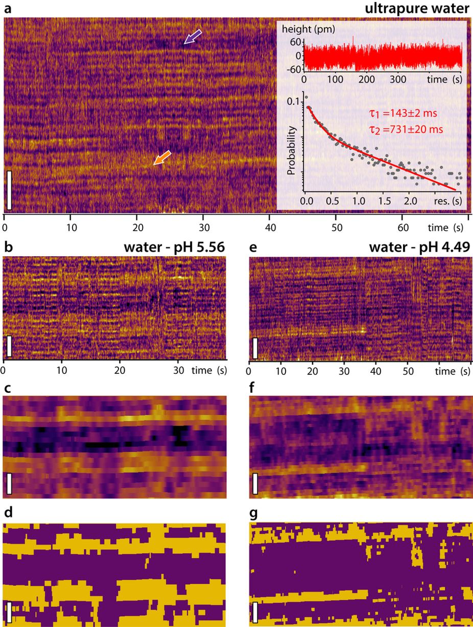
The dynamics of ions adsorbed at the surface of immersed charged solids plays a central role in countless natural and industrial processes such as crystal growth, heterogeneous catalysis, electrochemistry, or biological function. Electrokinetic measurements typically distinguish between a so-called Stern layer of ions and water molecules directly adsorbed on to the solid’s surface, and a diffuse layer of ions further away from the surface. Dynamics within the Stern layer remain poorly understood, largely owing to a lack of in-situ atomic-level insights. Here we follow the dynamics of single Rb+ and H3O+ ions at the surface of mica in water using high-resolution atomic force microscopy with 25 ms resolution. Our results suggest that single hydrated Rb+ions reside τ1 = 104 ± 5 ms at a given location, but this is dependent on the hydration state of the surface which evolves on a slower timescale of τ2 = 610 ± 30 ms depending on H3O+ adsorption. Increasing the liquid’s temperature from 5 °C to 65 °C predictably decreases the apparent glassiness of the interfacial water, but no clear effect on the ions’ dynamics was observed, indicating a diffusion-dominated process. These timescales are remarkably slow for individual monovalent ions and could have important implications for interfacial processes in electrolytes.
Maria Ricci, William Trewby, Clodomiro Cafolla, Kislon Voïtchovsky
Direct observation of the dynamics of single metal ions at the interface with solids in aqueous solutions
Nature Scientific Reports volume 7, Article number: 43234 (2017)
doi: https://doi.org/10.1038/srep43234
Please follow this external link for the full article: https://rdcu.be/4QVb
This article “Direct observation of the dynamics of sigle metal ions at the interface with solids in aqueous solutions” by Ricci, M. et al. is licensed under a Creative Commons Attribution 4.0 International License. The images or other third party material in this article are included in the article’s Creative Commons license, unless indicated otherwise in the credit line; if the material is not included under the Creative Commons license, users will need to obtain permission from the license holder to reproduce the material. To view a copy of this license, visit http://creativecommons.org/licenses/by/4.0/

![Figure 1 from "Ferroelectric Domain Studies of Patterned (001) BiFeO 3 by Angle- Resolved Piezoresponse Force Microscopy": Patterned mesas are separated from the continuous film by lithography, as shown in the AFM topography image. (b) Schematic drawing of the atomic structure of BFO with angle-resolved polarization models. The Fe (red sphere) atom can be displaced towards twelve possible polarization orientations with respect to its centrosymmetric position. (c) AR-PFM domain map of a 1.2 × 1.2 μm2 area of unpatterned BFO film, corresponding to the black dashed area in (a). (d) The area distribution of each polarization variant according to angle relative to the [100] direction. (e) The average area of stable and meta-stable polarization variants.](https://dhipgo7nn2tea.cloudfront.net/wp-content/uploads/2018/01/10172613/Figure_1_from_Ferroelectric_Domain_Studies.jpg)