Type: Arrow™ CONT

Contact Mode

Logo
Cantilever Data Value Range*
Resonance Frequency 14 kHz 10 - 19 kHz
Force Constant 0.2 N/m 0.06 - 0.38 N/m
Length 450 µm 445 - 455 µm
Mean Width 45 µm 40 - 50 µm
Thickness 2 µm 1.5 - 2.5 µm
ARROW™ AFM tip

ARROW™ AFM tip More images

Product Description

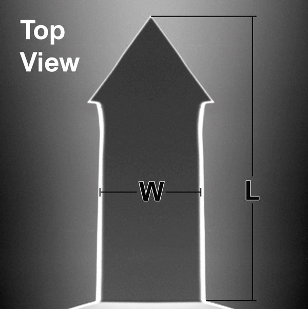
Optimized positioning through maximized AFM tip visibility

NanoWorld® Arrow™ CONT probes are designed for Contact Mode imaging. Furthermore this type can be used for Force Distance Spectroscopy Mode or Pulsed Force Mode (PFM). The CONT type is optimized for high sensitivity due to a low Force Constant.

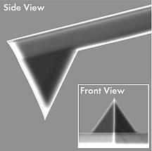
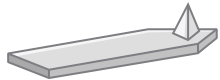
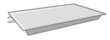
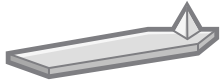
All SPM and AFM probes of the Arrow™ series are made from monolithic silicon which is highly doped to dissipate static charge. They are chemically inert and offer a high mechanical Q-factor for high sensitivity. These AFM probes feature a rectangular AFM cantilever with a triangular free end and a tetrahedral AFM tip with a typical height of 10 - 15 µm.

Additionally, this AFM probe offers a AFM tip radius of curvature of less than 10 nm.

The unique Arrow™ shape with the AFM tip position at the very end of the AFM cantilever allows easy positioning of the AFM tip on the area of interest.

Image 
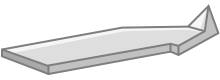
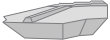
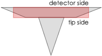
A trapezoidal cross section of the AFM cantilever and therefore 30% wider (e.g. NCH) AFM cantilever detector side result in easier and faster laser adjustment. Additionally, because there is simply more space to place and reflect the laser beam, a higher SUM signal is reached.

Tip shape: Arrow

Coating: none

Order Codes

Order Code Quantity Data Sheet
ARROW-CONT-10 10 Nominal values
ARROW-CONT-20 20 Nominal values
ARROW-CONT-50 50 Nominal values
ARROW-CONT-W 380 Nominal values

NanoWorld® Arrow™ Silicon AFM Probes Screencast

Subscribe to NanoWorld® Youtube Channel

Bruker® is a trademark of Bruker Corporation

Scientific publications mentioning use of this AFM probe


Piejko, Adrianna, Magdalena Tamulewicz‐Szwajkowska, Konrad Król, Karolina Ciesiołkiewicz, Robert Kudrawiec, and Jarosław Serafińczuk
Mechanical and Kelvin Probe Force Microscopy Investigations of Ultrathin Membranes Based on MoS2 and MoSe2
physica status solidi (b). 2024 Apr;261(4):2300352
DOI: https://doi.org/10.1002/pssb.202300352


Favrin, Fabio L., Lorenzo Zavagna, Matteo Sestini, Semih Esin, Bahareh Azimi, Massimiliano Labardi, Mario Milazzo, Giuseppe Gallone, Giovanna Batoni, and Serena Danti
Antifouling Properties of Electrospun Polymeric Coatings Induced by Controlled Surface Morphology
Energy & Environmental Materials. 2024 Nov;7(6):e12773
DOI: https://doi.org/10.1002/eem2.12773


Capaccio, Angela, Antonio Sasso, Oreste Tarallo, and Giulia Rusciano
Coral-like plasmonic probes for tip-enhanced Raman spectroscopy
Nanoscale. 2020;12(48):24376-84
DOI: https://doi.org/10.1039/D0NR05107A


Shao, Xueying, Maja Højvang Sørensen, Xingyu Xia, Chao Fang, Tsz Hin Hui, Raymond Chuen Chung Chang, Zhiqin Chu, and Yuan Lin
Beading of injured axons driven by tension-and adhesion-regulated membrane shape instability
Journal of the Royal Society Interface. 2020 Jul 29;17(168):20200331
DOI: https://doi.org/10.1098/rsif.2020.0331


Hesp NC, Torre I, Rodan-Legrain D, Novelli P, Cao Y, Carr S, Fang S, Stepanov P, Barcons-Ruiz D, Herzig-Sheinfux H, Watanabe K
Observation of interband collective excitations in twisted bilayer graphene
Nat. Phys. 17, 1162–1168 (2021)
DOI: https://doi.org/10.1038/s41567-021-01327-8


Genchi, Giada Graziana, Edoardo Sinibaldi, Luca Ceseracciu, Massimiliano Labardi, Attilio Marino, Sergio Marras, Giorgio De Simoni, Virgilio Mattoli, and Gianni Ciofani
Ultrasound-activated piezoelectric P (VDF-TrFE)/boron nitride nanotube composite films promote differentiation of human SaOS-2 osteoblast-like cells
Nanomedicine: Nanotechnology, Biology and Medicine. 2018 Oct 1;14(7):2421-32
DOI: https://doi.org/10.1016/j.nano.2017.05.006


Lau, Yu-Ting, Lam-Fung Kwok, Kin-Wai Tam, Ying-Shing Chan, Daisy Kwok-Yan Shum, and Graham Ka-Hon Shea
Genipin-treated chitosan nanofibers as a novel scaffold for nerve guidance channel design
Colloids and Surfaces B: Biointerfaces. 2018 Feb 1;162:126-34
DOI: https://doi.org/10.1016/j.colsurfb.2017.11.061


Becking, Jens, Albert Gröbmeyer, Martin Kolek, Uta Rodehorst, Susanne Schulze, Martin Winter, Peter Bieker, and Marian Cristian Stan
Lithium‐metal foil surface modification: an effective method to improve the cycling performance of lithium‐metal batteries
Advanced Materials Interfaces. 2017 Aug;4(16):1700166.
DOI: https://doi.org/10.1002/admi.201700166


Genchi, Giada Graziana, Luca Ceseracciu, Attilio Marino, Massimiliano Labardi, Sergio Marras, Francesca Pignatelli, Luca Bruschini, Virgilio Mattoli, and Gianni Ciofani
P (VDF‐TrFE)/BaTiO3 nanoparticle composite films mediate piezoelectric stimulation and promote differentiation of SH‐SY5Y neuroblastoma cells
Advanced healthcare materials. 2016 Jul;5(14):1808-20
DOI: https://doi.org/10.1002/adhm.201600245


Labuda, Aleksander, Marta Kocun, Martin Lysy, Tim Walsh, Jieh Meinhold, Tania Proksch, Waiman Meinhold, Caleb Anderson, and Roger Proksch
Calibration of higher eigenmodes of cantilevers
Review of Scientific Instruments. 2016 Jul 1;87(7)
DOI: https://doi.org/10.1063/1.4955122


Sarcevica, Inese, Liana Orola, Karol P. Nartowski, Yaroslav Z. Khimyak, Andrew N. Round, and László Fábián
Mechanistic and kinetic insight into spontaneous cocrystallization of isoniazid and benzoic acid
Molecular Pharmaceutics. 2015 Aug 3;12(8):2981-92
DOI: https://doi.org/10.1021/acs.molpharmaceut.5b00250


Kitta, Mitsunori, Toru Ide, Minako Hirano, Hiroyuki Tanaka, Toshio Yanagida, and Tomoji Kawai
Direct manipulation of a single potassium channel gate with an atomic force microscope probe
Small. 2011 Aug 22;7(16):2379-83
DOI: https://doi.org/10.1002/smll.201002337

For more information contact: info@nanoworld.com

Pointprobe® is a registered trademark of NanoWorld AG

All data are subject to change without notice.

NanoWorld AG
Rue des Saars 10
CH-2000 Neuchâtel,
Switzerland
www.nanoworld.com

For detailed information about our AFM probe product series please see below: